Phidolopora pacifica (Robertson, 1908)Common name(s): |
|
| Synonyms: Phidolopora labiata |  |
|
Class Gymnolaemata
Suborder Flustrina or
Ascophorina
Superfamily
Celleporoidea
Family
Phidoloporidae
|
|
| Phidolopora pacifica, collected subtidally from Sares Head. Total length 1.2 cm. | |
| (Photo by: Dave Cowles, June 2020) | |
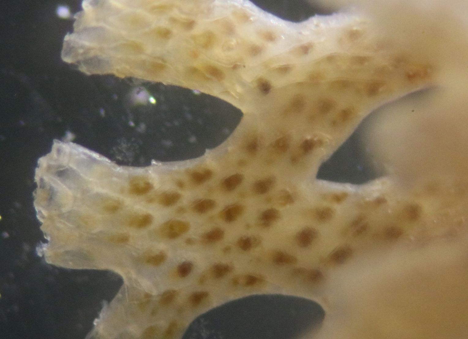
Description: As a member of Order Cheilostomatida, this species has box-shaped, calcified zooecia with opercula and often with spines. They also have several different forms of zooids including avicularia and vibracula. Embryos generally develop in brood chambers (ovicells).Phidolopora pacifica forms a unique, erect colony composed of a distinctive, lacy network of calcified material. The color is pale orange or salmon-orange when alive (as above).The primary aperture is symmetrical, sometimes with two distal spines and with the distal wall of the primary aperture beaded. There are avicularia on the frontal wall and large hooked avicularia on the dorsal side at the base of the fenestrations. All zooids open on the ventral side. The dorsal side of the colony is lined with kenozooids (membrane-coveres spaces without individual animals). The frontal walls have only a few small pores and few areolae.
How to Distinguish from Similar Species:Phidolopora labiata is also a valid species, but according to marinespecies.org what was formerly called P. labiata in the Pacific Ocean is now called Phidolopora pacifica.
Geographical Range: Pacific ocean, with subspecies names for forms apparently from Catalina California and Japan.
Depth Range:
Habitat:
Biology/Natural
History: This form
of "chicken wire"-like fenestrated network structure in bryozoans is
referred
to as "retroporid".
| Return to: | |||
| Main Page | Alphabetic Index | Systematic Index | Glossary |
References:
Dichotomous Keys:Carlton, 2007
Kozloff, 1987, 1996 (as Phidolopora labiata)
General References:
Scientific Articles:
Ayer, Stephen W., Raymond J. Andersen, Cunheng He, and Jon Clardy,
1984. Phidolopine, a new purine derivative from the bryozoan Phidolopora
pacifica. The Journal of Organic Chemistry 49 20 pp
3869-3870 https://doi.ort/10.1021/jo00194a053
Tischler, Mark, Stephen W. Ayer, and Raymond J. Andersen, 1986. Nitrophenols from northeast Pacific bryozoans. Comparative Biochemistry and Physiology part B: Comparative biochemistry 84:1 pp 43-45
Web sites:
General Notes and Observations: Locations, abundances, unusual behaviors:
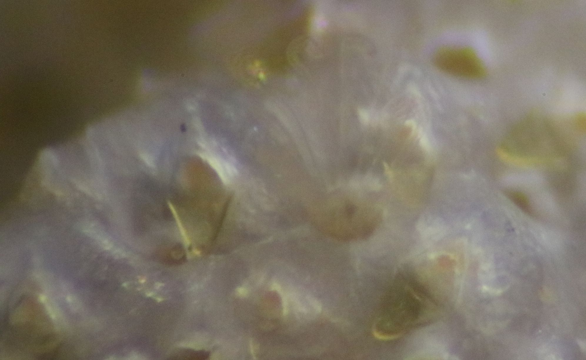
A closeup of the colony showing one open lophophore,
plus rounded opercula
and several pointed avicularia.
All the zooids
appear
to be facing outward on the colony. This view of the back of one of the
fenestrations, facing the inside of the colony, shows a similar overall
structure as the front but with no individual zooids.
Therefore this side shown must be the dorsal side which has kenozooids
(membrane-covered areas) instead of individual zooids.
Authors and Editors
of Page:
Dave Cowles (2018): Created original page
CSS coding for page developed by Jonathan Cowles
Salish Sea Invertebrates web site provided courtesy of Walla
Walla University