Tritonia tetraquetra Bergh, 1894Common name(s): Pink tritonia, Diomedes tritonia |
|
| Synonyms: Duvaucelia gilberti, Sphaerostoma diomedea, Tritonia diomedea (part), Tritonia exsulans | 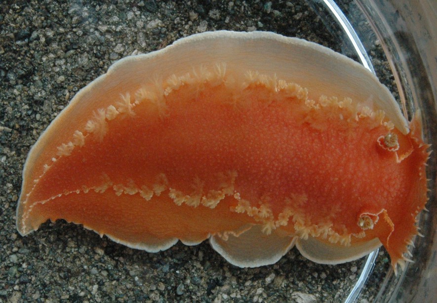 |
|
Class Gastropoda
Family Tritoniidae
|
|
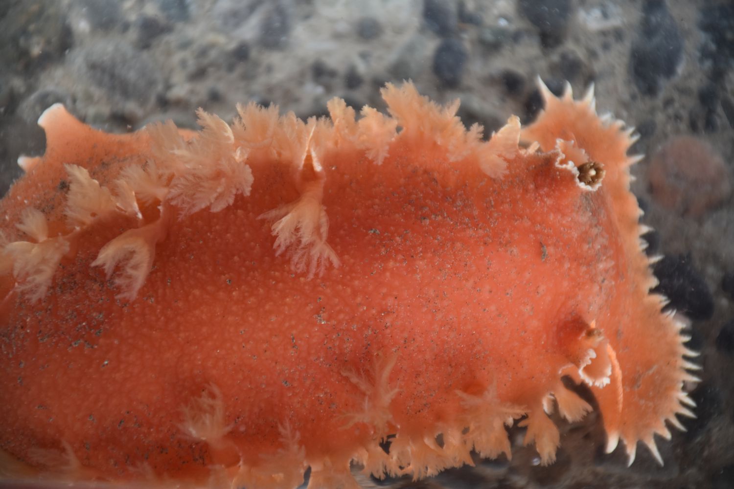
| Tritonia tetraquetra, about 12 cm long, collected near the mouth of Bowman Bay. | |
| (Photo by: Dave Cowles, August 2014 ) | |
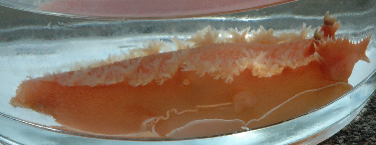
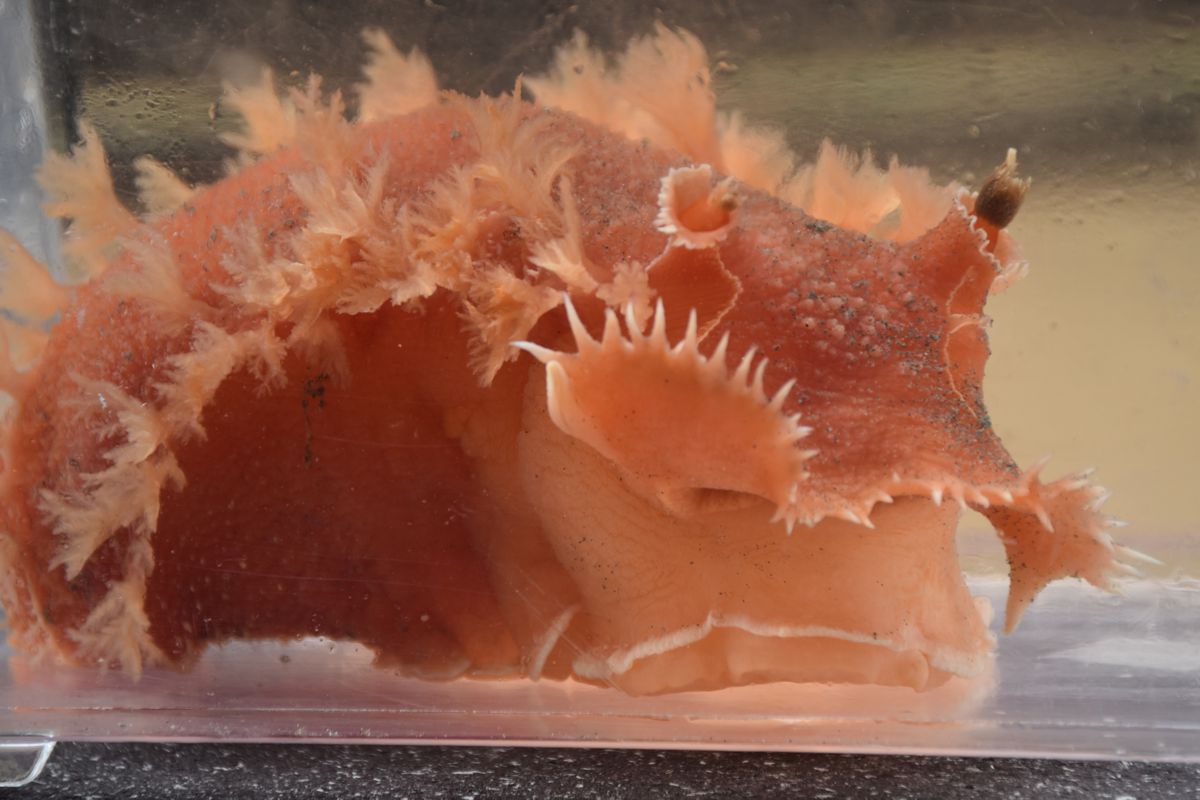
Description: As with other dendronotid nudibranchs, the anus is on the right side of the body instead of dorsal or posterior (photo). The clavus of the rhinophores can be withdrawn into the sheath (photo). There are bushy or paddle-like cerata or gills on the dorsum, usually in longitudinal rows (photo). Tritonia diomedia has a massive body, longer than wide and wider than high, and a longitudinal row of gills along a definite dorsolateral margin on each side of the smooth dorsum. The 20-30 gill tufts in each row are separated from one another by spaces. The oral veil has about 18 (10-30) short, simple (unbranched) white papillae. Color is salmon or rose without a pattern of white lines, except that there is a narrow white line along the margin of the dorsum, the margin of the foot, and the edge of the rhinophore sheath. The edge of the rhinophere sheath is smooth but the white-tipped clavus of the rhinophore is surrounded by about 20 yellow to brownish processes (photo). Length up to 22 cm.
How to Distinguish from Similar Species:Tochuina gigantea, which also eats sea pens, has unbroken rows of white gill tufts along the margins of the dorsum. Tritonia festiva is smaller and has a pink dorsum, often with a pattern of white lines. Tritonia exsulans is difficult to distinguish but it is smaller, has a body about as wide as high and several differences in internal anatomy
Geographical Range: In the north Pacific from Japan through Alaska down to Panama. Also in Florida.
Depth Range: Intertidal to 656 m (mostly subtidal)
Habitat: On sandy bottoms, especially near sea pens.
Biology/Natural History:
Feeds
on sea pens such as Ptilosarcus
gurneyi, Stylatula
elongata,
and Virgularia
sp.
| Return to: | |||
| Main Page | Alphabetic Index | Systematic Index | Glossary |
References:
Dichotomous Keys:Carlton, 2007
Kozloff, 1987, 1996
General References:
Behrens, 1991
Behrens and Hermosillo, 2005
Harbo, 2011
Lamb and Hanby, 2005
Scientific Articles:
Web sites:
Dany Burgess blog on the Washington Department of Ecology page for Feb 14, 2022
General Notes and Observations: Locations, abundances, unusual behaviors:
This view of the right side of the animal clearly shows the anus, which
opens on a papilla
at about the middle of the right side. The gonopore,
here releasing a string of eggs, is forward of the anus. Note
also
the unusual height of the dorsum.
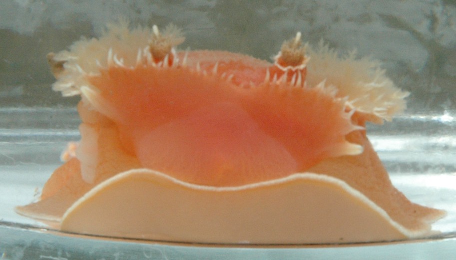
This view of the face shows the white line bordering the foot; the
10-30 white papillae
along the oral
veil, and the white line and distinctive brown processes
surrounding
the clavus of the rhinophore.
The gonopore
is visible to the left, releasing a chain of eggs.
This view of another individual nearly head-on shows how high the body often is.
A dorsal view. Notice that the rhinophores are almost totally withdrawn into the sheath. Photo by Dave Cowles, July 2018
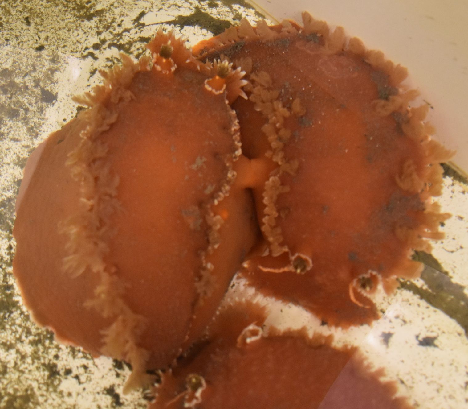
Since
the gonopore is located on the right side, individuals copulate by
lining up head-to-tail with right sides facing. A third individual is
investigating at the bottom of the photo. Photo by Dave Cowles, July
2018.
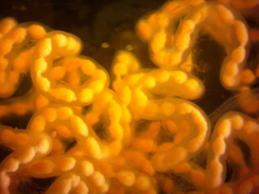
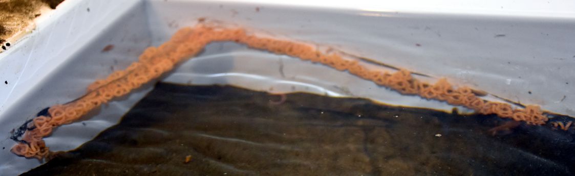
The eggs are orange, about 1 mm long, and laid in a loose string.
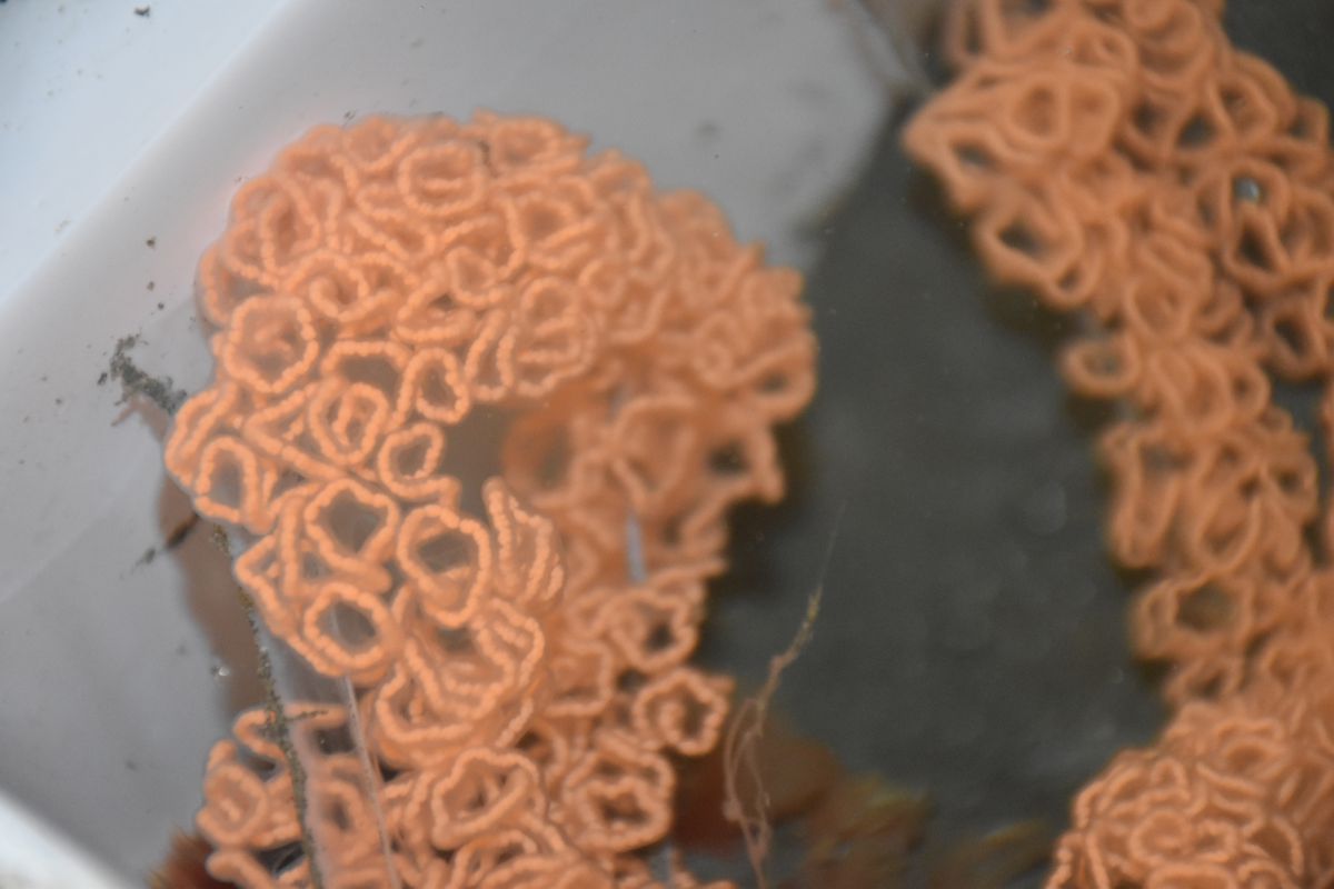
The eggs may be deposited in clusters (above) or strings (below).
Authors and Editors of
Page:
Dave Cowles (2014): Created original page
CSS coding for page developed by Jonathan Cowles (2007)
Salish Sea Invertebrates web site provided courtesy of Walla
Walla University