Hydractinia milleri Torrey, 1902Common name(s): Miller hydractinia, Hedgehog hydroid, Snail fur |
|
| Synonyms: | 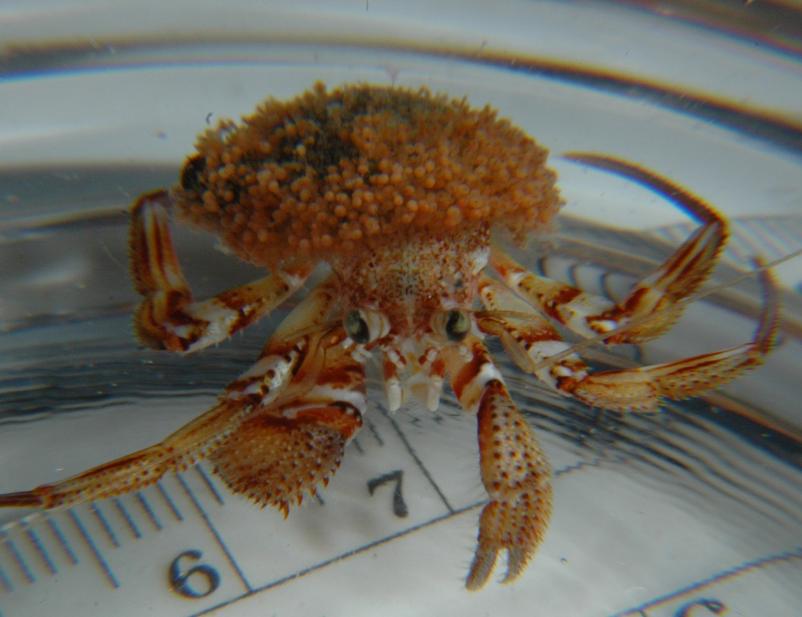 |
|
Phylum Cnidaria
Class Hydrozoa
Suborder Athecata
(Anthomedusae)
Family Hydractiniidae
|
|
| Hydractinia milleri forming a "house" on the subtidal hermit crab Pagurus dalli. Total diameter of "house" is 2 cm. Specimen was captured at 20 m at Mukilteo. | |
| (Photo by: Dave Cowles, Dec 2007) | |
How to Distinguish from Similar Species: All Hydractinia species grow in a similar mat of stolons covered with perisarc, with naked (no perisarc), unbranched polyps arising individually from the mat. H. laevispina and an unnamed local Hydractinia species have 8 tentacles. H. laevispina has pink gastrozooids and many short, curved spines on the perisarc while the unnamed Hydractinia has few, long, uncurved spines in the mat and the gastrozooids are white except near the mouth. H. aggregata has 20-24 tentacles on the gastrozooids and females carry several eggs. Several of the Hydractinia species can be found growing on hermit crab shells.
Note: Some gastrozooid polyps of this colony seem to have only 8 tentacles, which would make it H. laevispina. Others have 12 or more tentacles, however. It could perhaps be a hybrid between H. laevispina and H milleri.
Geographical Range: Vancouver Island, Canada to Monterey Bay, CA
Depth Range: Low intertidal and subtidal.
Habitat: Sides and undersides of rocks and on hermit crab shells
Biology/Natural History:Hydractinia species are often found on the shells of hermit crabs, though I can find no report of Hydractinia living on this species of hermit crab (Pagurus dalli), which usually inhabits sponges. Colonies if this species bear only one sex of gonozooids--the above colony is bearing females. The gonozooids produce medusoids which are little more than gonads and are not released from the colony as free-living medusae. In related Hydractinia species, release of eggs and sperm is light-dependent and occurs in the morning. Newly settled invidividuals of H. milleri have found in August in Monterey Bay. The hermit crab seems to frequently rub the colony with the flagellum of its second antennae. In hermit crab symbioses with related species of Hydractinia, this action has been shown to result in the hermit crab's scraping off some of the larger plankton captured on the Hydractinia and provides a supplementary food source for the hermit crab. Thy hydroid consumes a variety of small planktonic species such as crustacean larvae, nematodes, and even small benthic animals. Predators of Hydractinia include nudibranchs such as Dendronotus and Cuthona spp. If two colonies of Hydractinia occur on the same shell they seem to remain 1-2 mm apart from one another.
The presence of this hydroid on a hermit crab seems to at least partially deter predation by octopus. Octopus usually readily capture hermit crabs and other crustaceans. However, an octopus clearly thinks twice about attacking a hermit crab with Hydractinia on its shell. Click here for a movie showing how octopus deal with Hydractinia-covered hermit crabs.Hydractinia colonies are complex and consist of 4 types of polyps. The colonies are either male or female, and shed gametes into the water. After fertilization, a planula larva develops. The planula settles on a hermit crab shell, crawls around, and metamorphoses into a polyp. This first polyp (a gastrozooid, shaped like a typical polyp with tentacles and a gastrovascular cavity) begans to sprout ropelike stolons from its base. The stolons are hollow and continuous with the gastrovascular cavity, ectoderm, and gastrodermis. These stolons spread across the shell, gripping the shell surfacee, and begin to grow up periodically into other polyps (also gastrozooids). The stolon network becomes more and more interconnected, then the stolons begin to widen into a flattened mat. This mat connects all the polyps and is innervated in its upper layer. After the stolon mat has covered the entire gastropod shell the hermit crab is living in, several new types of polyps begin to grow. Reproductive gonozooids arise from the mat. These polyps have gonophores (where gametes are made) sticking out from their column, and don't have well-developed tentacles. Dactylozooids are specialized, usually smaller finger-like polyps which only grow along the aperture of the shell the hermit crab is living in. The dactylozooids seem to specialize in capturing hermit crab eggs. Tentaculozooids are quite long and tentacle-like, and about as large as an entire gastrozooid polyp. They grow in various areas of the colony and are used for defense (information from Cartwright, 2003)
As a side note, Hydractinia spp (not this species but especially H. echinata and H. symbiolongicarpus) have long been used as model organisms for animal development (Gahan et al., 2016). Hydractinia colonies have two components: anemone-like polyps and pipe-like stolons, which connect the different polyps and attach to the substrate (photo). The first, settled animal is a single polyp which begins to grow stolons which asexually bud new polyps from them. Its tissues contain cells called "interstitial cells" (called "i-cells for short), especially in the epidermis. I-cells are small cells which reside in the interstitial spaces among regular epithelial cells. They have a large nucleus but not much cytoplasm and divide frequently, as well as migrate. The suite of genes they contain is similar to that seen in stem cells and germ cells of more complex organisms. I-cells are in both polyps and stolons, but in polyps they mostly cluster near the base of the column while in stolons they are scattered more randomly. If a polyp is injured the i-cells rapidly collect there and seem to be the main agents in regeneration. They do the same in stolons but more slowly. It is currently not clear whether the i-cells are multipotent (or even totipotent), or whether they consist of morphologically indistinguishable lines of cells which together regenerate the different cell types of the colony. I-cells express several genes characteristic of stem cells and several others characteristic of specific cell lines, such as neural cells. Proliferation of the cells, formation of the oral end of the polyp, and commitment to neural cell lineage is associated with Wnt signaling. Wnt signaling needs to be inhibited in order for cells to the stolon, and if Wnt expression is inhibited, the polyps become stolons. Hydractinia can regenerate any lost part, and, like the frewhwater Hydra, do not appear to have age-related decline (increase in mortality or lowered reproductive potential). In other words, Hydractinia seem to enjoy immortal youth!
| Return to: | |||
| Main Page | Alphabetic Index | Systematic Index | Glossary |
References:
Dichotomous Keys:Flora and Fairbanks, 1966 (as Hydractinia sp)
Kozloff 1987, 1996
Smith and Carlton, 1975 (as Hydractinia sp)
General References:
American
Fisheries Society, 2002
Harbo,
1999
Morris
et al., 1980
Ricketts
et al., 1985
Scientific Articles:
Cartwright, Paulyn, 2003.
Developmental insights
into the origin of complex colonial hydrozoans. Integrative and
Comparative Biology 43: pp 82-86
Web sites:
General Notes and
Observations: Locations,
abundances, unusual behaviors:
I do not often see hermit crab houses made of Hydractinia, as in this case, and I have never found a Pagurus dalli hermit crab with Hydractinia. On this individual the greatest density of polyps is near the edges of the "house" close to the hermit crab, while most of the "house" distant from the hermit crab is composed of the stolon mat with a few scattered polyps. The most common hermit crab species that I have found in this area to have Hydractinia "houses" are deeper-living Labidochirus splendescens.
My thanks to Kirt Onthank for his sharp-eyed viewing during a dive which resulted in finding this symbiotic pair.
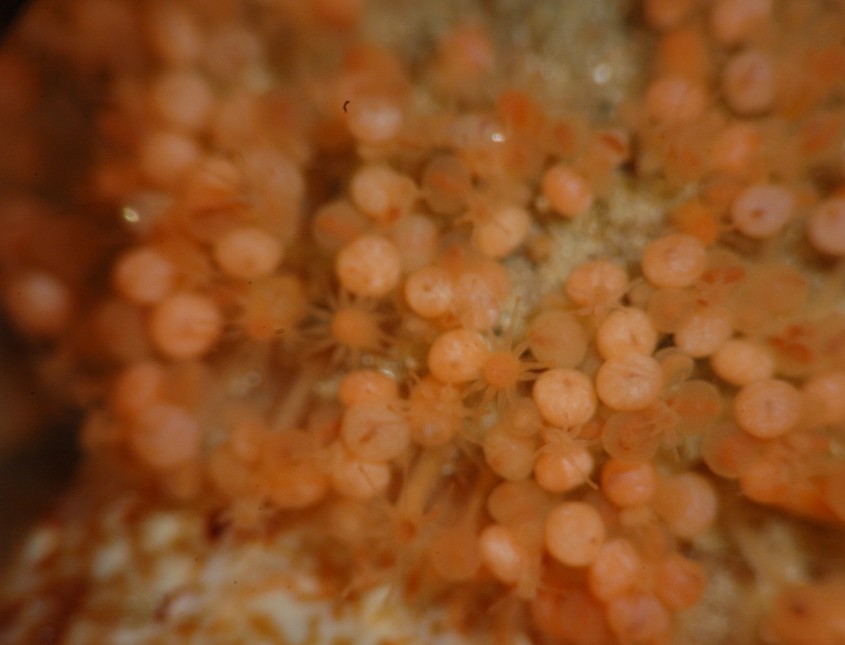
This closeup shows details of the colony morphology. The
matlike
meshwork of stolons
covered with perisarc
can be seen here and there between the polyps
(the stippled color at the bottom left is the carapace of the hermit
crab
host that the hydroids are forming a "house" for). Individual
polyps,
not covered by perisarc,
arise from the mat. The feeding gastrozooids
are pink and have around 8 tentacles (some seem to have
more). The
female reproductive gonozooids
(called gonophores
when there are carrying eggs) carry a single egg which mostly fills the
gonozooid.
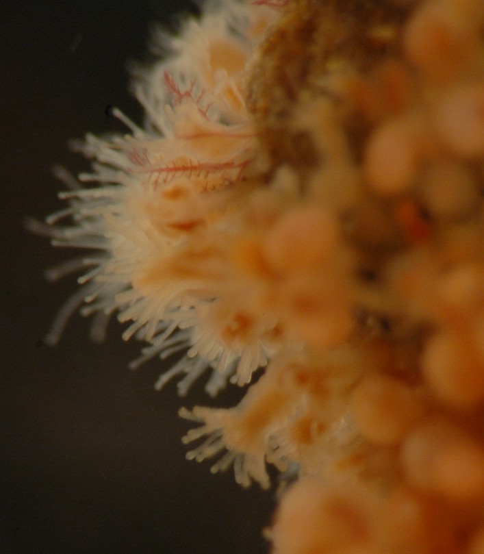
This closeup of the edge of the colony shows the gastrozooids.
A few tiny stalks of a red alga seem to have begun growth within the
colony.
Authors and Editors of Page:
Dave Cowles (2007): Created original page
CSS coding for page developed by Jonathan Cowles (2007)