Hydractinia laevispina Fraser, 1922Common name(s): Snail fur hydroid |
|
| Synonyms: | 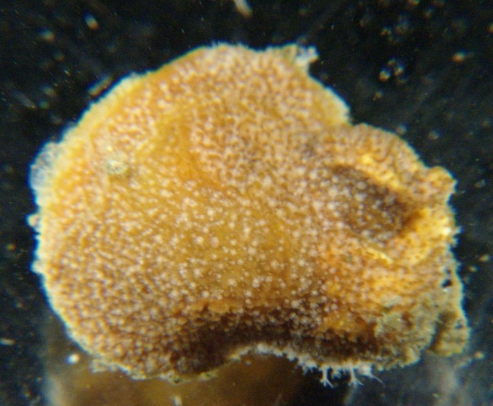 |
|
Phylum Cnidaria
Class Hydrozoa
Suborder Athecata
(Anthomedusae)
Family
Hydractiniidae
|
|
| Hydractinia laevispina growing as an extension to a small gastropod shell inhabited by the hermit crab Labidochirus splendescens. Collected from 60-100 m depth in the San Juan Channel, July 2010. The shell is to the bottom right and the extension which has grown beyond the shell is to the upper left. Total colony width about 2 cm. | |
| (Photo by: Dave Cowles ) | |
How to Distinguish from Similar Species: All Hydractinia species grow in a similar mat of stolons covered with perisarc, with naked (no perisarc), unbranched polyps arising individually from the mat. Hydractinia sp has 8 tentacles but the hypostome of the gastrozooids is white and the mat has fewer, longer spines. Gastrozooids of Hydractinia milleri and H. aggregata have 12-24 tentacles.
Geographical Range: In portions of the the Pacific and Arctic oceans.
Depth Range:
Habitat: Grows on snail shells inhabited by hermit crabs.
Biology/Natural History: Predators on Hydractiniahydroids include the nudibranchs Dendronotus frondosus and Cuthona divae.
This colony was inhabited by a hermit crab, Labidochirus splendescens. The crab's long legs extended far beyond the limits of the colony and could not be even partially drawn inside. When the crab was presented to a hungry red octopus, Octopus rubescens, the octopus quickly pulled the hermit crab out of the shell, dropped the shell with the hydroid colony, and ate the crab.
Hydractinia colonies are complex and consist of 4 types of polyps. The colonies are either male or female, and shed gametes into the water. After fertilization, a planula larva develops. The planula settles on a hermit crab shell, crawls around, and metamorphoses into a polyp. This first polyp (a gastrozooid, shaped like a typical polyp with tentacles and a gastrovascular cavity) begans to sprout ropelike stolons from its base. The stolons are hollow and continuous with the gastrovascular cavity, ectoderm, and gastrodermis. These stolons spread across the shell, gripping the shell surfacee, and begin to grow up periodically into other polyps (also gastrozooids). The stolon network becomes more and more interconnected, then the stolons begin to widen into a flattened mat. This mat connects all the polyps and is innervated in its upper layer. After the stolon mat has covered the entire gastropod shell the hermit crab is living in, several new types of polyps begin to grow. Reproductive gonozooids arise from the mat. These polyps have gonophores (where gametes are made) sticking out from their column, and don't have well-developed tentacles. Dactylozooids are specialized, usually smaller finger-like polyps which only grow along the aperture of the shell the hermit crab is living in. The dactylozooids seem to specialize in capturing hermit crab eggs. Tentaculozooids are quite long and tentacle-like, and about as large as an entire gastrozooid polyp. They grow in various areas of the colony and are used for defense (information from Cartwright, 2003)
As a side note, Hydractinia
spp (not this species but especially H. echinata and H. symbiolongicarpus)
have long been used as model organisms for animal development
(Gahan et al., 2016). Hydractinia
colonies have two components: anemone-like polyps
and pipe-like stolons,
which connect the different polyps and
attach to the substrate (photo).
The first, settled animal is a single
polyp
which begins to grow stolons
which asexually bud new polyps
from
them. Its tissues contain cells called "interstitial cells" (called
"i-cells for short), especially in the epidermis. I-cells are small
cells which reside in the interstitial spaces among regular epithelial
cells. They have a large nucleus but not much cytoplasm and divide
frequently, as well as migrate. The suite of genes they contain is
similar to that seen in stem cells and germ cells of more
complex organisms. I-cells are in both polyps
and stolons,
but in
polyps
they mostly cluster near the base of the column while in stolons
they are scattered more randomly. If a polyp
is injured the i-cells
rapidly collect there and seem to be the main agents in regeneration.
They do the same in stolons
but more slowly. It is currently not clear
whether the i-cells are multipotent (or even totipotent), or whether
they consist of morphologically indistinguishable lines of cells which
together regenerate the different cell types of the colony. I-cells
express several genes characteristic of stem cells and several others
characteristic of specific cell lines, such as neural cells.
Proliferation of the cells, formation of the oral end of the polyp,
and
commitment to neural cell lineage is associated with Wnt signaling. Wnt
signaling needs to be inhibited in order for cells to the
stolon,
and if Wnt expression is inhibited, the polyps
become stolons.
Hydractinia
can
regenerate any lost part, and, like the frewhwater Hydra, do not
appear
to have age-related decline (increase in mortality or lowered
reproductive
potential). In other words, Hydractinia
seem to enjoy immortal youth!
| Return to: | |||
| Main Page | Alphabetic Index | Systematic Index | Glossary |
References:
Dichotomous Keys:Carlton, 2007
Kozloff, 1987, 1996
General References:
American
Fisheries Society, 2002
Morris
et al., 1980
Scientific Articles:
Cartwright, Paulyn, 2003. Developmental insights
into
the origin of complex colonial hydrozoans. Integrative and
Comparative
Biology 43: pp 82-86
Gahan, James M., Brian Bradshaw, Hakima Flici, and Uri Frank, 2016. The interstitial stem cells in Hydractinia and their role in regeneration. Current Opinion in Genetics & Development 40: pp65-73. doi 2443/10/j.gde.2016.06.006
Web sites:
General Notes and
Observations: Locations,
abundances, unusual behaviors:
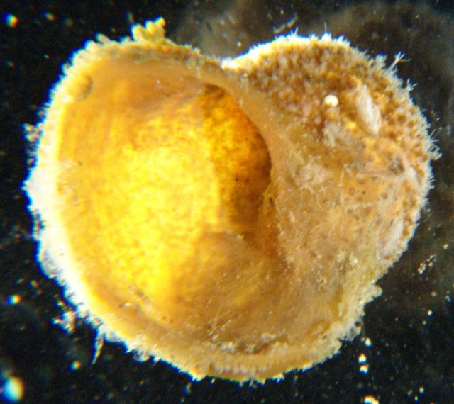
This view shows the underside of the colony. The original
shell
the colony encrusted is to the top right and the expansion which has
grown
beyond the shell, which covered the hermit crab's carapace, is to the
left.
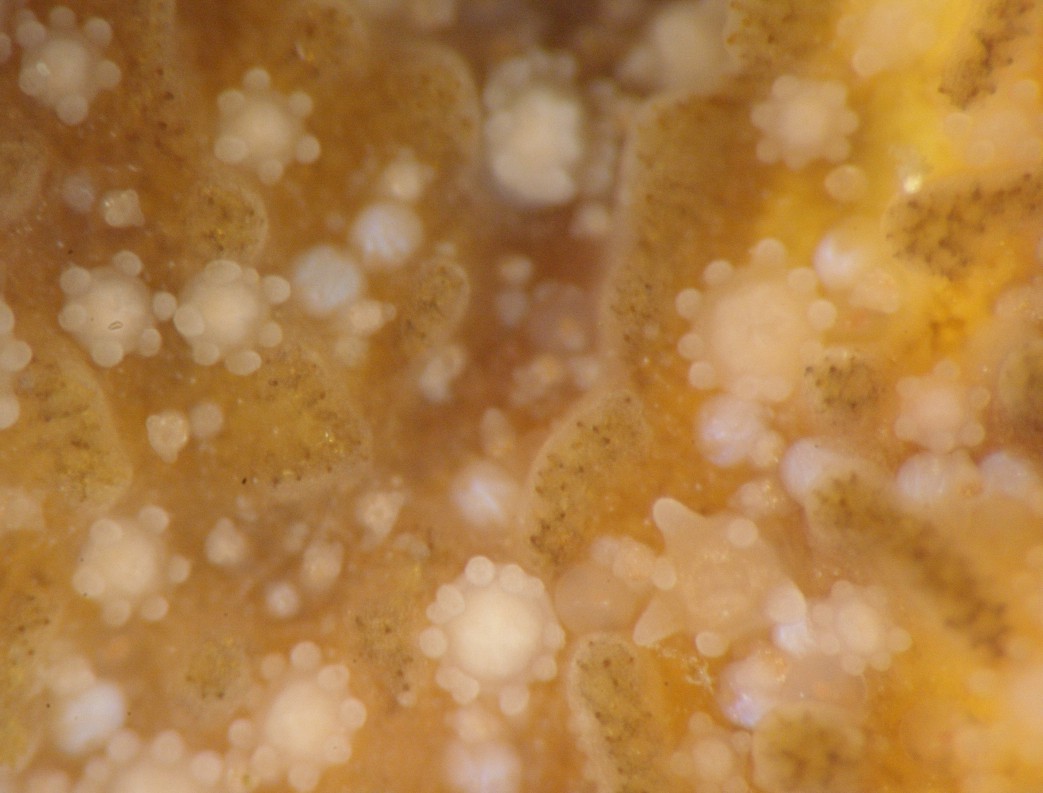
This closeup view shows partly expanded polyps, which are about 0.5
mm diameter. Several of these polyps
have more than 8 tentacles. Note also the blunt, short,
slightly
curved spines arising from the stolon
mat between the polyps.
A few of the smallest polyps
may be dactylozooids.
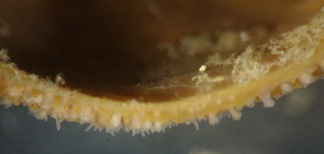
This view of the edge of the colony shows how the stolons
weave together to make a solid mat.
Authors and Editors of Page:
Dave Cowles (2010): Created original page
CSS coding for page developed by Jonathan Cowles (2007)
Rosario Invertebrates web site provided courtesy of Walla Walla University