Oplophorus spinosus Brulle'Common name(s): |
|
| Synonyms: | 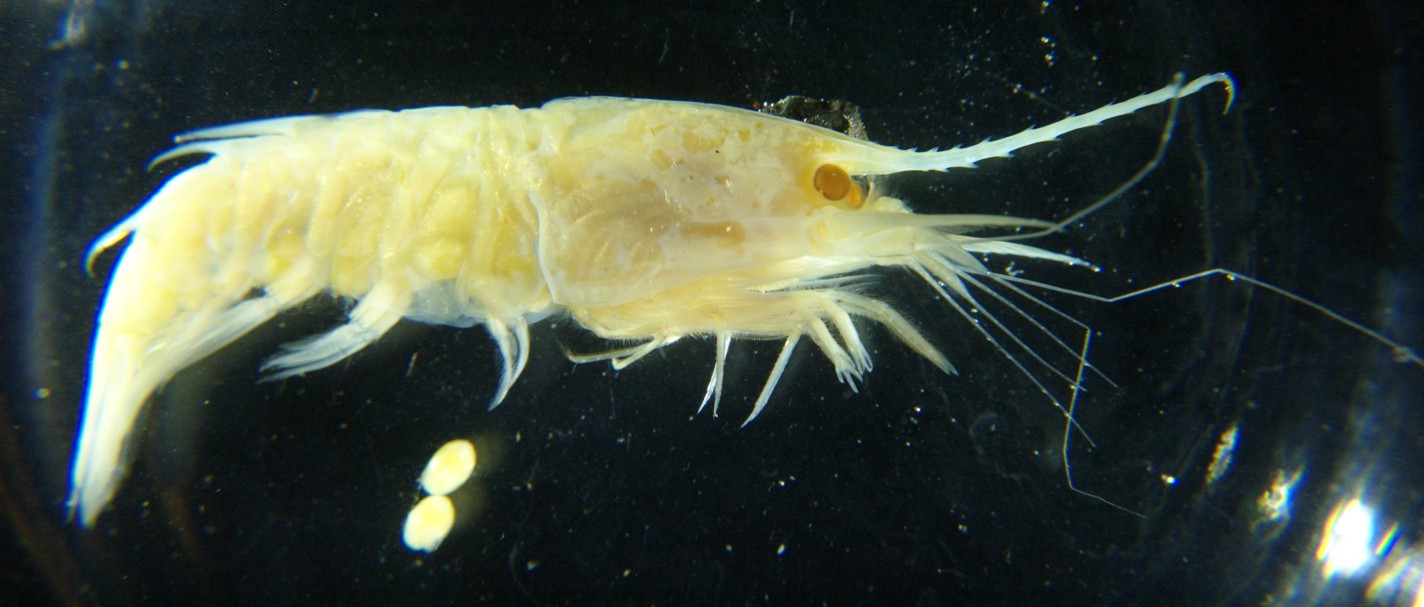 |
|
Phylum Arthropoda
Subphylum Crustacea
Class Malacostraca
Subclass Eumalacostraca
Superorder Eucarida
Order Decapoda
Suborder Pleocyemata
|
|
| A female Oplophorus spinosus carrying eggs, carapace length 1.5 cm, captured in midwater off Hawaii in 1996. In real life this species would be partly pinkish-red and partly transparent. The specimen above has been preserved in formalin. Several eggs have dropped off the pleopods. | |
| (Photo by: Dave Cowles, 2012 ) | |
Description: This is a true shrimp from the family Oplophoridae, which lives in deep midwater offshore. True (Caridean) shrimp have the second abdominalepimera overlapping that of segment 1 and 2. Family Oplophoridae is almost entirely midwater, has exopodites on its pereopods, and pereopods 1 and 2 are longer and more stout than the others. Members of genus Oplophorus have a general surface that is hard and polished. The rostrum has as many or more dorsal teeth as ventral (photo). The eyes are at least as wide as the eyestalk (photo). They have a well-developed epipod on the 4th pereopod. The abdomen has no posterior mid-dorsal spine on the 2nd segment (photo) but the 5th abdominal segment has a posterior mid-dorsal tooth (which may be small) (photo), and the 5th segment is longer than the 6th segment (photo). The 6th abdominal segment is not carinate on the dorsal side (photo). The telson is pointed posteriorly but does not end with a spinose endpiece (photo). The appendix masculina on the 2nd abdominalpleopod of males is much longer than the appendix interna. Oplophorus females have few (less than 50), relatively large eggs (photo). Oplophorus spinosus has no sharp tooth along the posterior end of the ventral margin of the carapace in adults (photo). The antennal scale has a barb on the inner (mesial) margin not far fron the anterior end (this is the origin of the species' name) (photo) and teeth along the lateral margin (photo).
How to Distinguish from Similar Species: O. gracilirostris and O. typus have a sharp tooth near the posteror end of the ventral margin of the carapace. O. novaezeelandiae does not but it also does not have the distinctive backward-pointing spine on the inner margin of the antennal scale. Once individuals have been identified asOplophorus, they can be readily identified to this species by running a fingernail along the inner margin of the antennal scale to feel for the spine.
Geographical Range: The Indian Ocean, In the Pacific off Japan, Hawaii, Easter Island, and a number of seamounts, subtropical Atlantic.
Depth Range: Mesopelagic. Up to as shallow as 140 m at night; down as far as 750 m during the day.
Habitat: Mesopelagic
Biology/Natural History:
This species
is a very fast swimmer and a daily vertical migrator. The carapace
of most Oplophorus
species
I have examined is laterally compressed more than in most other shrimp
species. Most deep-sea species have eyes sensitive mainly to
blue
light since that is the predominant color at depth. Frank
and Widder (1996) report that this species is one of several
that also
has eyes sensitive to ultraviolet light, and that they can likely
detect
UV light downwelling from the surface at their normal daytime
depth.
This species also has several ventral
photophores that can be rotated so that they continue to direct light
downward
while the animal changes position (Nowel
et al., 1998), probably to help the animal obliterate its
shadow.
The luciferin used by this species may be derived from Coelenterazine
(produced
by Cnidarians)
from its diet. Promega has developed a product 'Nanoluc
Luciferase'
based on the luciferase of a closely related species, O.
gracilirostris.
Nanoluc luciferase is a smaller, brighter molecule than most other
luciferases
such as firefly luciferase and GFP, which helps optimize it for cases
of
tracking low gene expression.
| Return to: | |||
| Main Page | Alphabetic Index | Systematic Index | Glossary |
References:
Dichotomous Keys:Chace, Fenner A., Jr., 1986. The Caridean shrimps (Crustacea: Decapoda) of the Albatross Philippine expedition, 1907-1910, part 4: Families Oplophoridae and Nematocarcinidae. Smithsonian Contributions to Zoology Nol 432. 82 pp.
General References:
Scientific Articles:
Cowles, D.L., 1987. Factors affecting the aerobic
metabolism
of midwater crustaceans. Ph.D. dissertation, University of
California,
Santa Barbara. 228 pp.
Cowles, D.L., J.J. Childress, and M.E. Wells,
1991. Metabolic
rates of midwater crustaceans as a function of depth of occurrence off
the Hawaiian Islands: food availability as a selective
factor? Marine
Biology 110: pp 75-83
Frank,
Tamara M. and Edith A. Widder, 1996. UV light in
the deep-sea:
In situ measurements of downwelling irradiance in relation to the
visual
threshold sensitivity of UV-sensitive crustaceans. Marine and
Freshwater
Behavior and Physiology 27: No. 2-3 pp 189-197
Gaten,
Edward, and Peter J. Herring, 2005. Morphology of
the reflecting
superposition eyes of larval oplophorid shrimps. Journal of
Morphology
225:1 pp 19-29
Gaten,
E., P.M.J. Shelton, and P.J. Herring, 1992.
Regional morphological
variations in the compound eyes of certain mesopelagic shrimps in
relation
to their habitat. Journal of the Marine Biological
Association of
the United Kingdom 72: Issue 01 pp 61-75
Holthuis, L.B., 1949. The caridean crustacea of
the Canary
Islands. Zoologische mededelingen uitgegeven door het
Rijksmuseum
van natuurlijke histoie te Leiden XXX: No. 15.
Nowel,
M.S., P.M.J. Shelton, and P.J. Herring, 1998.
Cuticular photophores
of two decapod crustaceans, Oplophorus spinosus and
Systellaspis
debilis. Biological Bulletin 195: pp 290-307
Shelton,
P.M.J., E. Gaten, and P.J.Herring, 1992.
Adaptations of tapeta
in the eyes of mesopelagic decapod shrimps to match the oceanic
irradiance
distribution. Journal of the Marine Biological Association of
the
United Kingdom 72: Issue 01 pp 77-88
Web sites:
General Notes and Observations: Locations, abundances, unusual behaviors:
The photos below are of a preserved specimen captured off Hawaii in 1996. In real life much of the body would be translucent or transparent while parts of the thorax are colored light pink.
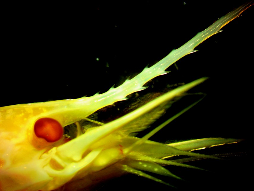
As with other Oplophorus species, O. spinosus has a long rostrum with many spines on both the dorsal and ventral surface. Note the well-developed eye as well.
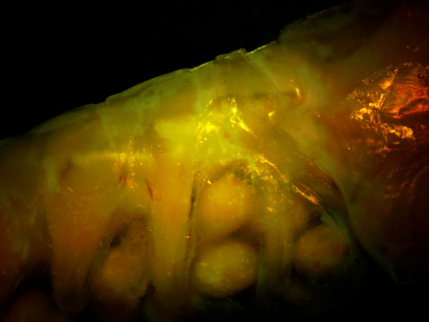
This right-side view of the anterior (first 3) abdominal segments (the posterior thorax is at the right side) shows that the second abdominal segment does not have a large posterior mid-dorsal spine. The large eggs can be seen attached to the pleopods.
This right-side view of the posterior abdominal segments show the large posterior-pointing mid-dorsal spines on several of them. Abdominal segment 3 is at the top right, then segments 4,5,6, and the base of the telson is visible at the bottom left. Note that segment 6 is not dorsally carinate.
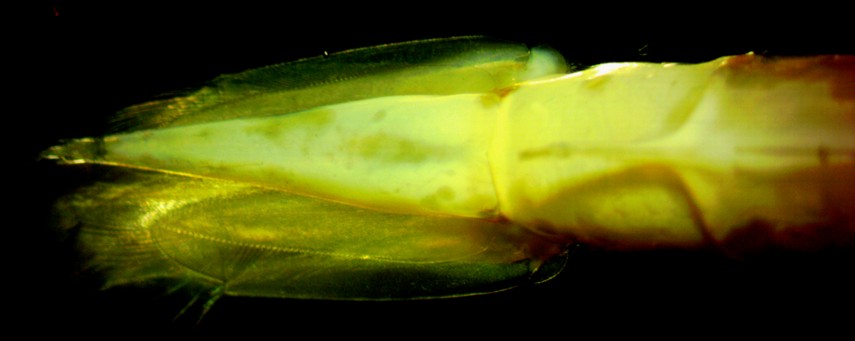
Dorsal view of the rear abdomen and telson. The rightmost segment is abdominal segment 5. Segment 6 is below the posterior spine from segment 5. To the left are the uropods and telson.
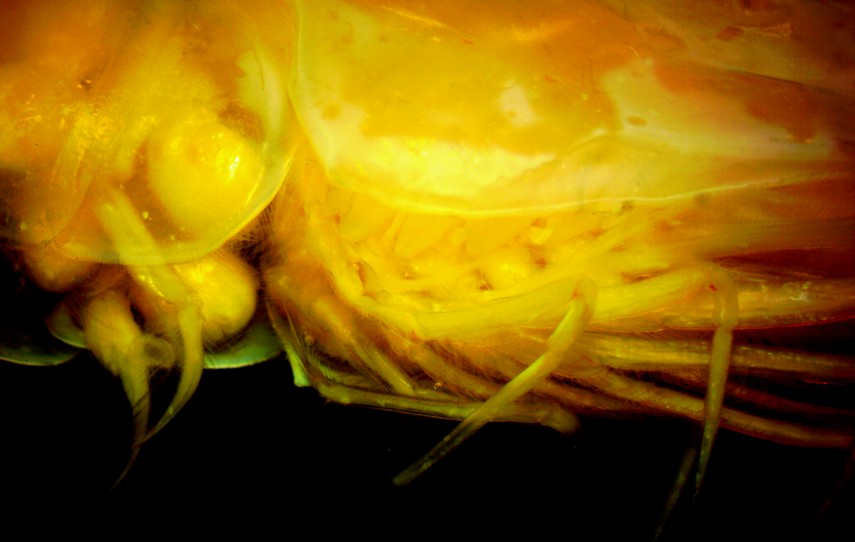
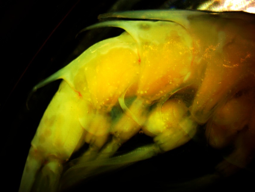
In O. spinosus there is no sharp tooth on the posterior margin of the ventralcarapace. In this view of the animal's right side, the ventral margin of the carapace plus the pereopods can be seen to the right and the first abdominal segment with pleopods and several eggs is visible to the left. The posterior margin of the ventralcarapace has an acutely rounded corner but no sharp tooth.
 |
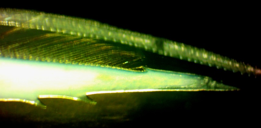 |
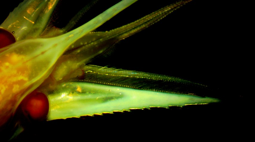
| These views show the characteristic pattern of teeth and spines on the antennal scale. The left view is a dorsal view of the head, showing the rostrum, eye, antennae, antennules, and antennal scale. Notice the numerous forward-pointing teeth on the outer (lateral) margin of the antennal scale, while the inner (mesial) margin has a backward-pointing spine near the anterior end and setae along much of its margin. The right view is a closeup of the end of the right antennal scale, showing the distinct backward-pointing spine on the mesial margin near the anterior end. | |
Authors and Editors of Page:
Dave Cowles (2012): Created original page
CSS coding for page developed by Jonathan Cowles (2007)
Salish Sea Invertebrates web site provided courtesy of Walla Walla University