Pagurus stevensae Hart, 1971Common name(s): Steven's hermit crab |
|
| Synonyms: Pagurus brandtii | 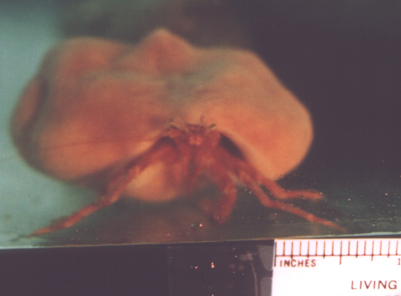 |
| Phylum Arthropoda
Subphylum Crustacea Class Malacostraca Subclass Eumalacostraca Superorder Eucarida Order Decapoda Suborder Pleocyemata Infraorder Anomura Family Paguridae |
|
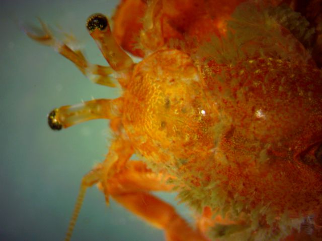
| Pagurus stevensae captured at 80 m depth in the San Juan Channel | |
| (Photo by: Dave Cowles July 2001) | |
How to Distinguish from Similar Species: P. dalli has only one row of spines on the dorsal surface of its left chela and the carpus of the right cheliped is only about 1.5x as long as wide, plus it has a distinct white band on the distal part of the merus of the chelipeds.
Geographical Range: Akun Island(Bering Sea) to Puget Sound
Depth Range: Subtidal, 5-198 m
Habitat: On shell/gravel bottoms.
Biology/Natural
History:
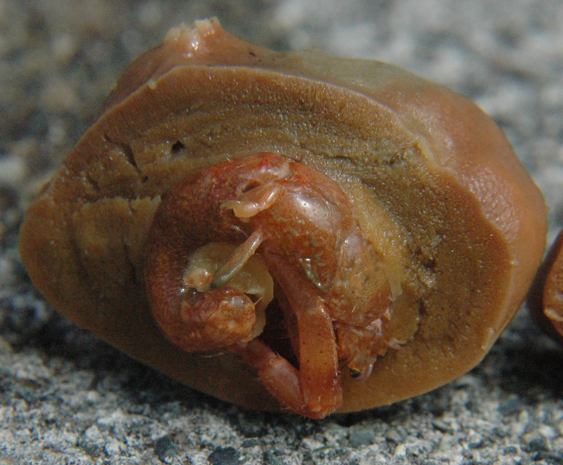
This individual
is living in the sponge Suberites
suberea forma latus.
It may actually be inside the
fragments of a badly eroded shell which the sponge covered.
This
species usually lives in sponges, though it may live in a shell as
well.
| Return to: | |||
| Main Page | Alphabetic Index | Systematic Index | Glossary |
References:
Dichotomous Keys:Hart, 1982
Kozloff 1987, 1996
General References:
Harbo,
1999
Jensen,
1995
Scientific
Articles:
Hart 1971, J. Fish. Res. Board Can. 28(10):1537. (original description)
General Notes and Observations: Locations, abundances, unusual behaviors:
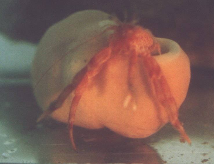
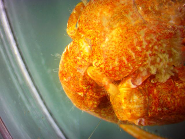
Another photo of the same individual as above.
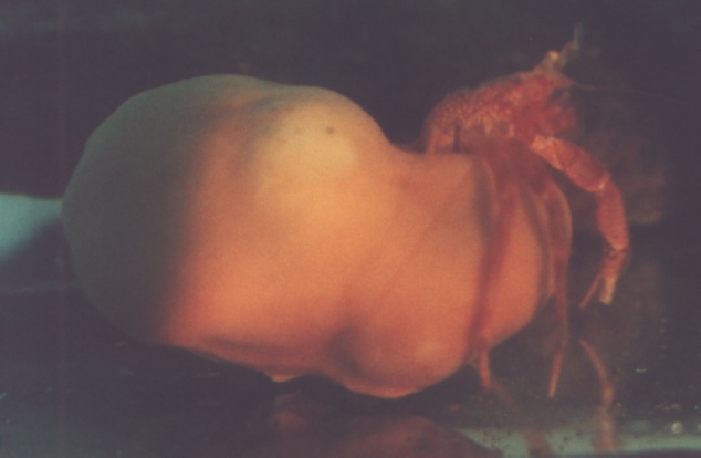
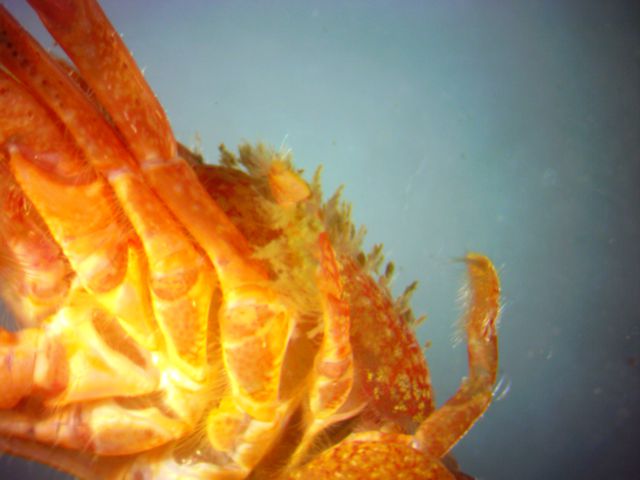
The same individual trying to right the sponge.
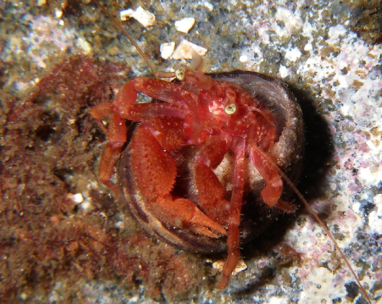
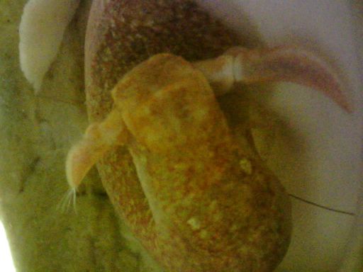
An underwater photo of Pagurus stevensae by Aaron
Baldwin
Guide to keying this species in Kozloff's key: Photos by Dave Cowles, August 2016, of an animal captured at 100 m depth in the San Juan Channel
The carapace
is smooth and only the carapace
shield is calcified. The basal portions of the eyescales
are not covered by the carapace.
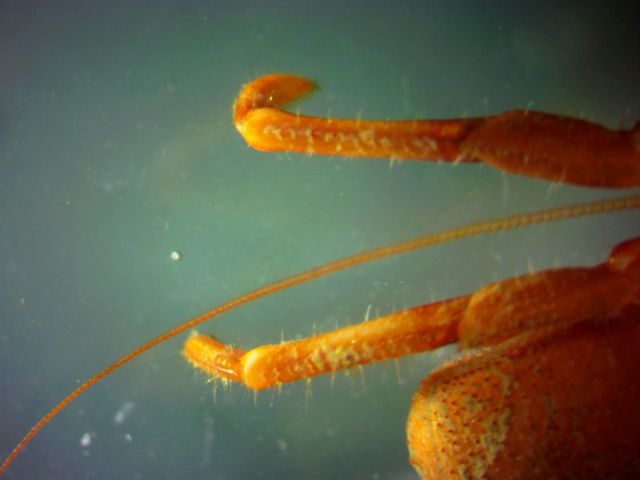
The eyescales
do not have a deep median
groove and they end with only one point. The rostrum
is inconspicuous and not pointed.
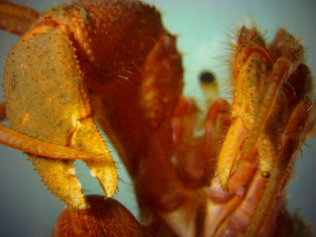
Leg 4, shown here in dorsal
view on the right side of an animal out of its shell, is reduced in
size
and subchelate.
Here is a ventral
view of all 5 legs on the left side of the animal. Note that
both
legs 4 and 5 are reduced in size.
The uropods are asymmetrical (one longer than the other)
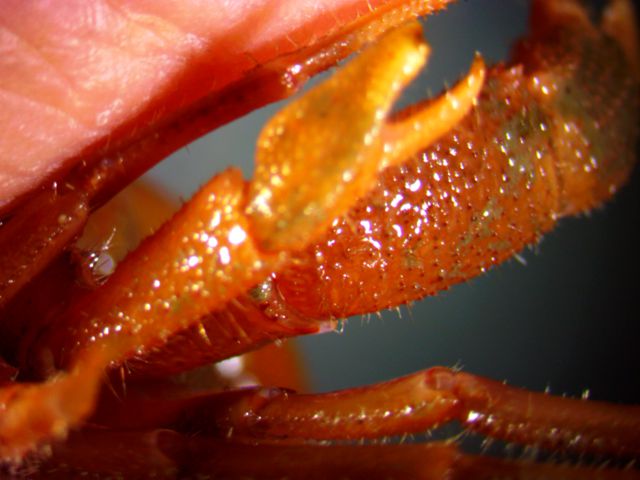
The carpus
of the right cheliped
is about twice as long as wide.
The dactyls
of legs 2 and 3 are not obviously twisted in relation to the propodus,
and do not have reddish stripes. The antenna is not striped.
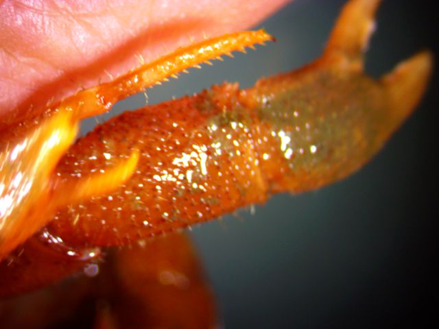
The dorsal
surface of the palm (propodus)
of the left chela
does not have a prominent ridge or crest near the midline. It
does,
however, have two rows of spines.
The right chela
is larger than the left. (Since this is a ventral
view, the right chela
is on the left and the left chela
is on the right)
The ventral
side of the merus
of the right cheliped
(visible to the lower right) has several small spines or tubercles,
but does not have two tubercles
which are markedly larger than the rest.
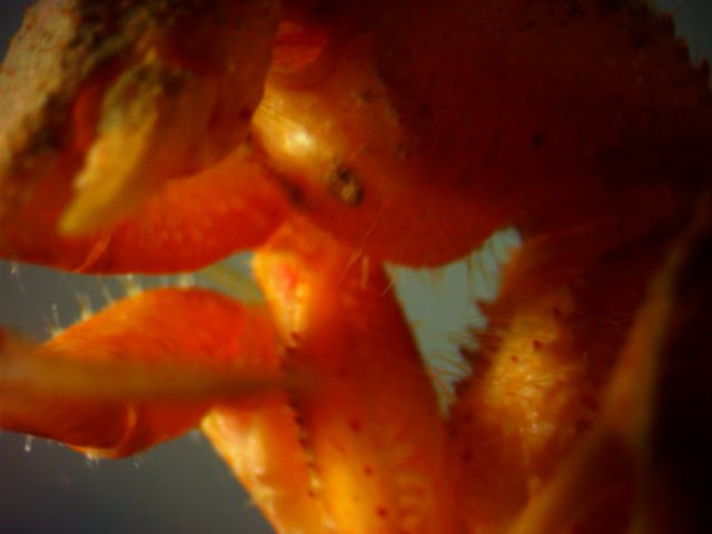
The dactyl
of the left chela
closes firmly against the propodus
without a major gap between. Here, although the propodus
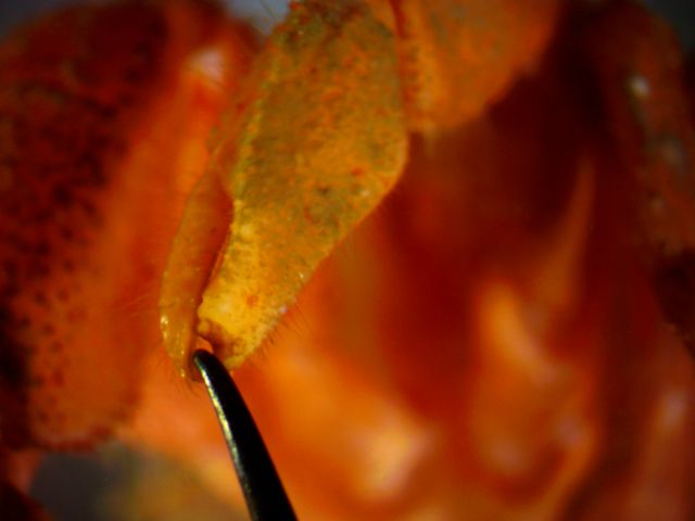
is slightly damaged, the crab is tightly gripping my probe.
The carpus
of the left cheliped
is about 3x as long as wide. The left chela
is approximately triangular when closed.
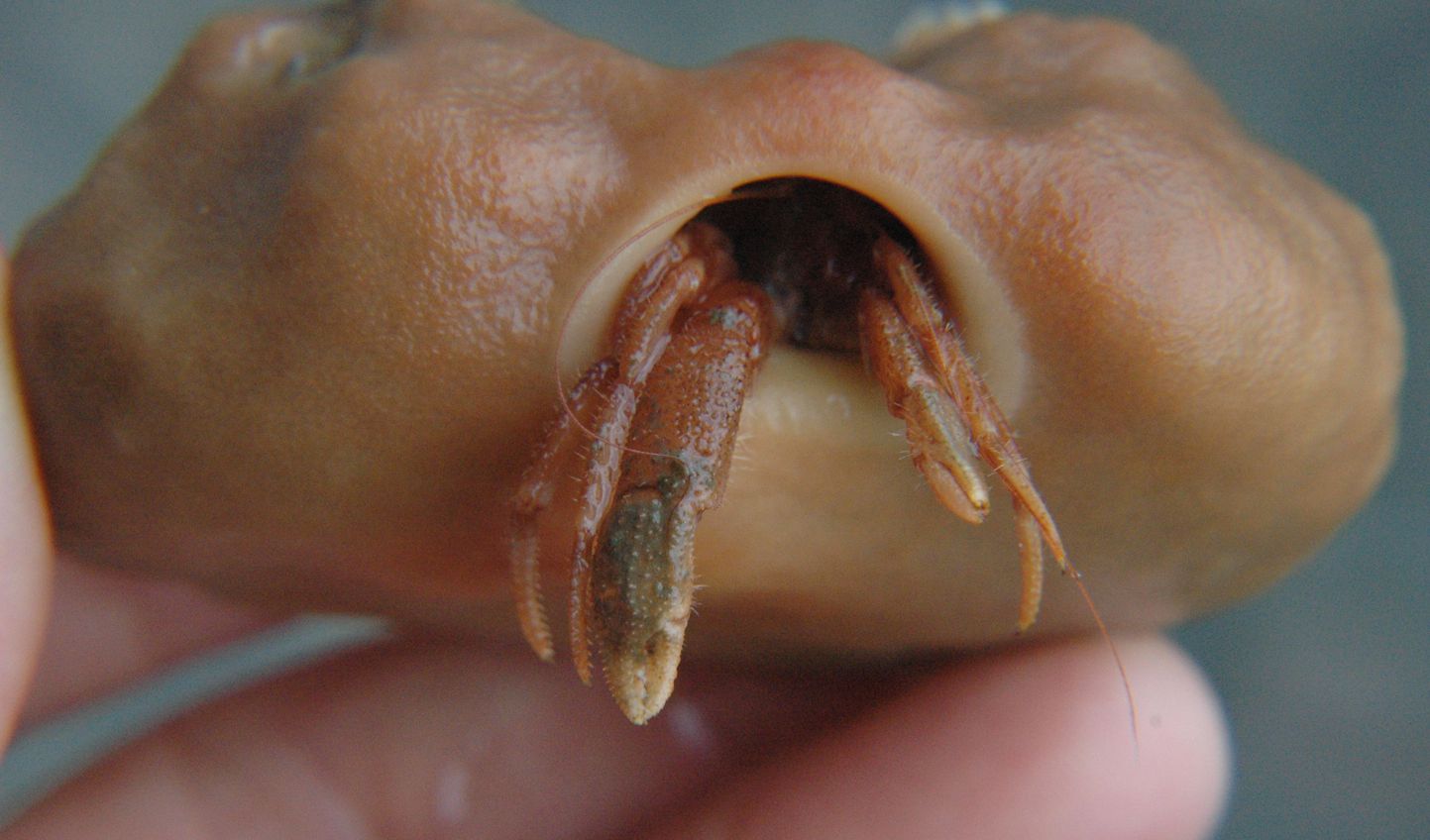
This frontal view of the live hermit crab in its Suberites
suberea
forma latus
sponge shows that there is no white band on the merus
of either cheliped.
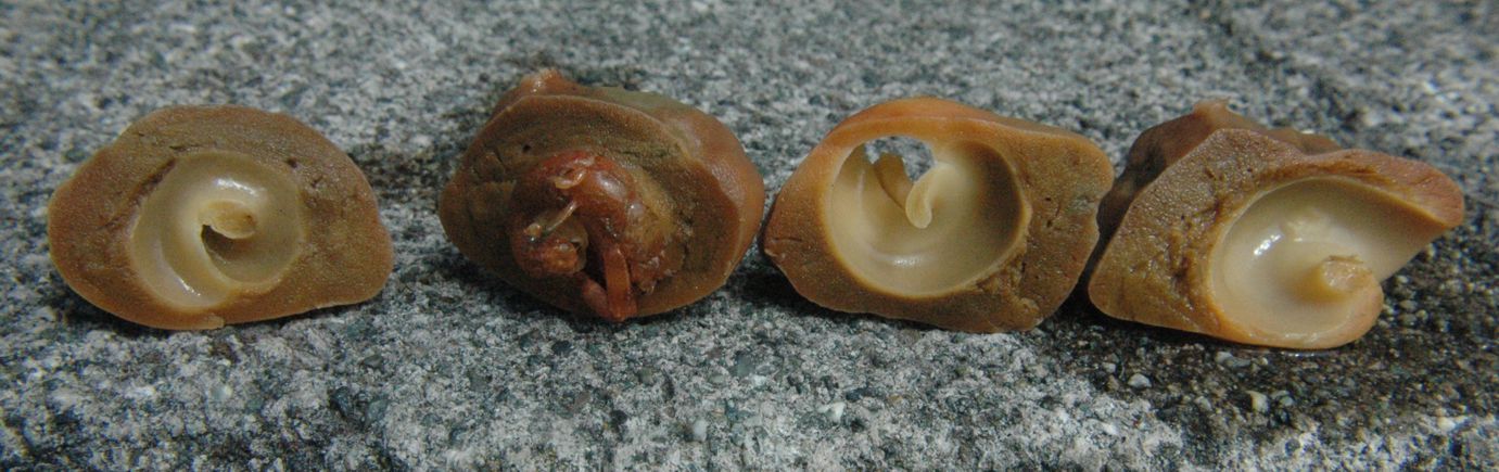
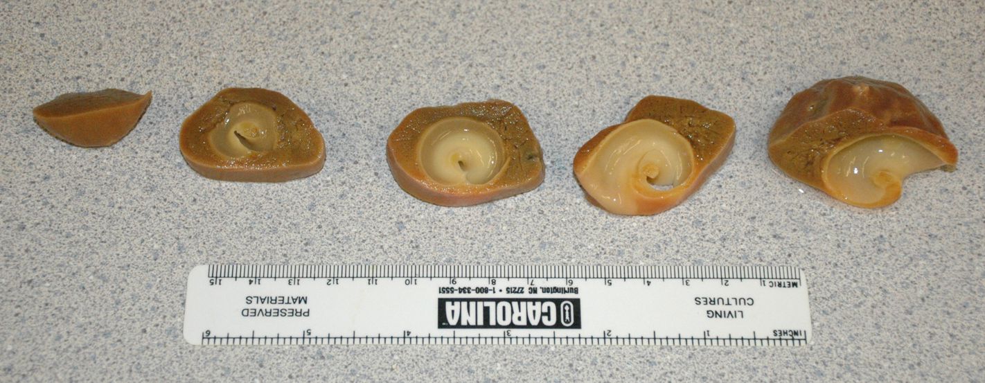
| I cut open the Suberites suberea sponge from the above individual to see how large a residual shell remained inside. I could find no trace of a shell left--all the passages were purely of sponge. It looked like the passageway involved at least 4-5 loops. The hermit crab retreated into the sponge and clung to it tightly as I carved it away. Don't worry--I didn't harm the hermit crab and I gave it a nice, roomy shell to live in when I was finished, which it picked up and packed off. |
 |
 |
 |
Authors and Editors of Page:
Dave Cowles (2005): Created original page