Cranopsis cucullata (Gould, 1846)Common name(s): Hood puncturella, Hooded puncturella, Helmet puncturella |
|
| Synonyms: Puncturella cucullata | |
|
Class Gastropoda
Order Archaegastropoda
Suborder Pleurotomariina
|
|
| Cranopsis cucullata collected from a red octopus (Octopus rubescens) midden. Length 1 cm. | |
| (Photo by: Dave Cowles March 2008) | |
How to Distinguish from Similar Species: Puncturella. multistriata has about 30 primary ribs, each separated by 1-3 secondary ribs. The primary ribs do not project more than 1 mm beyond the shell margin. Puncturella galeata has no anterior seam, about 49-63 ribs, and only grows to 2 cm. Other keyhole limpets such as Diodora aspera and Fissurellidea bimaculata have a round opening at the apex instead of a slit in front.
Geographical Range: Alaska to Cabo San Qintin, Baja California
Depth Range: Low intertidal to 200 m; more common subtidally
Habitat: On and under rocks
Biology/Natural History:
Cranopsis was formerly considered to be a subset of Puncturella.
| Return to: | |||
| Main Page | Alphabetic Index | Systematic Index | Glossary |
References:
Dichotomous Keys:Flora and Fairbanks, 1967 (as P. cucullata)
Kozloff 1987, 1996
General References:
American
Fisheries Society, 1998
Harbo,
1997
Morris,
1966 (as P.
cucullata)
Sept,
1999
Scientific Articles:
Web sites:
General Notes and Observations: Locations, abundances, unusual behaviors:
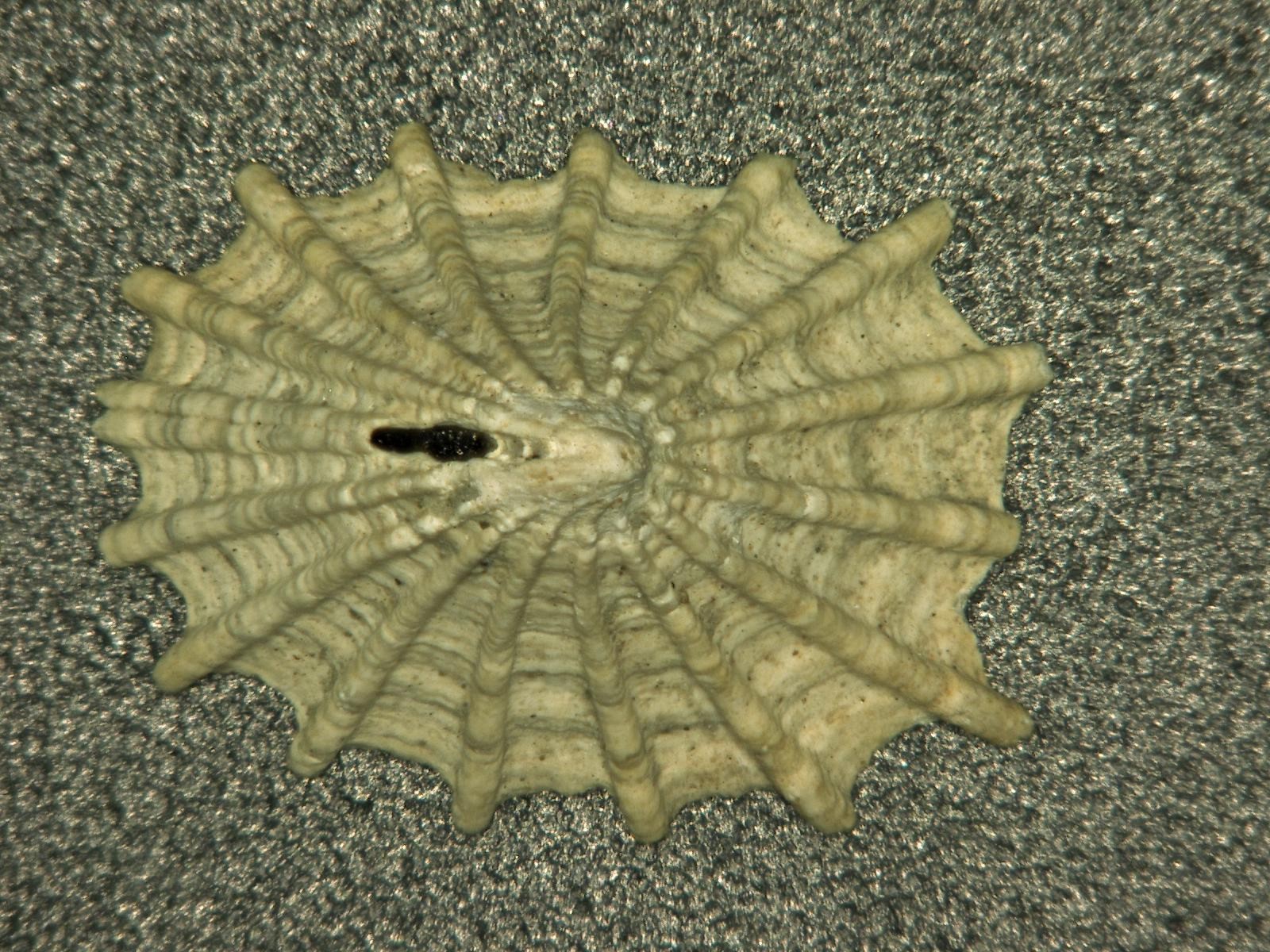
This closeup shows the anterior slit and the seam which leads from the slit to the anterior edge of the shell. This photo was made from a stack of photos which were combined by a computer algorithm to create a much greater depth of field (photo by Dave Cowles, technology by Keyence DHX-100 digital microscope and software)
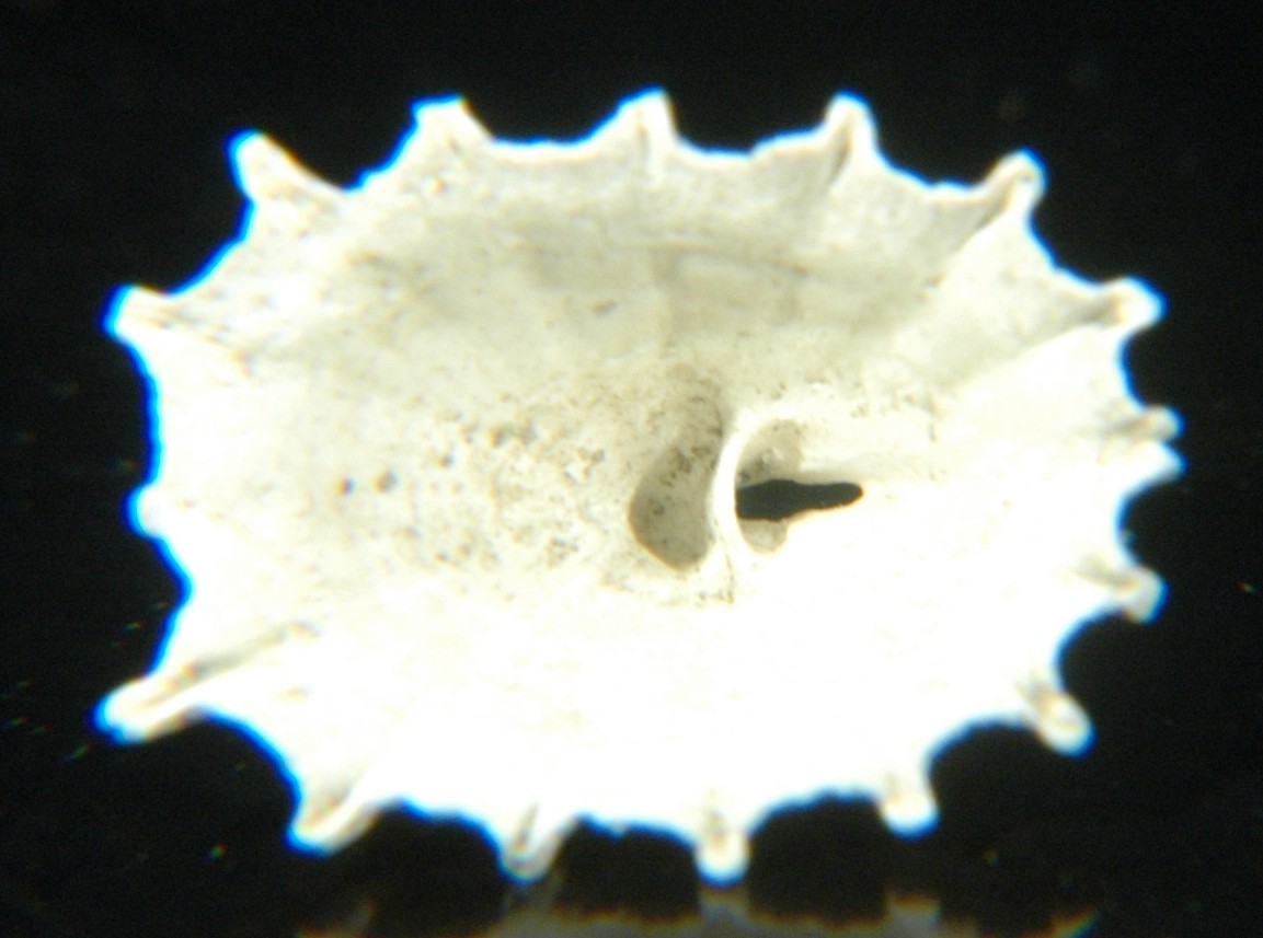
The anterior slit is separated from the actual apex of the shell by
a partition, as can be seen in this view of the underside.
Authors and Editors of Page:
Dave Cowles (2008): Created original page
CSS coding for page developed by Jonathan Cowles (2007)