Aeolidia loui Keinberger et al., 2016Common name(s): Shag-rug aeolis, shag rug nudibranch, mossy nudibranch, shaggy mouse nudibranch, common grey sea slug, maned nudibranch, papillose aeolid, warty sea mouse |
|
| Synonyms: Was earlier identified as A. papillosa | 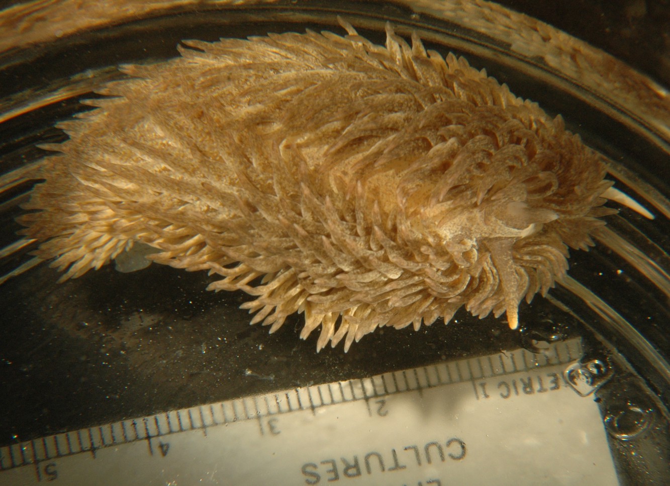 |
|
Class Gastropoda
Suborder Aeolidacea
|
|
| Aeolidia loui, approximately 4.5 cm long, found on a rock in Padilla Bay. This individual is crawling around the side of a dish. The rhinophores are visible in front with light tips, while one white pedal tentacle is visible on the extreme right. | |
| (Photo by: Dave Cowles, July 2008) | |
How to Distinguish from Similar Species: Specimens in our area were all formally recognized (and keyed in Kozloff 1987, 1996) as A. papillosa but that species is now recognized as part of a species complex (Kienberger et al., 2016). Molecular and morphological studies have identified two species present here in the Salish Sea: Aeolidia papillosa and Aeolidia loui. Aeolidia papillosa has smooth rhinophores and cerata which remain approximately the same width from base to near the top and ranges mainly north from here to Alaska, over to Japan, and in the Atlantic, while A. loui has "warty" rhinophores and (sometimes bristly) cerata which are wider at the base than at the tips and ranges from British Columbia south to California. This difference in rhinophores may be the most useful morphological feature for distinguishing the two species here while live, although the rhinophores may become smooth after preservation. Flabellina salmonacea looks similar but no cerata are attached anterior to the rhinophores. A deep-water species, A. herculea, lives at depths greater than 500 m.
Geographical Range: Northeast Pacific, from British Columbia to California
Depth Range: Intertidal to 900 m
Habitat: On rocks, or may be on floats or docks. Often near its perferred prey, Anthopleura elegantissima.
Biology/Natural History: Feeds on anemones, especially Anthopleuraelegantissima and secondarily Metridium senile. Also may feed on Urticina crassicornis, Anthopleura xanthogrammica, and Epiactis prolifera, the young of which it may swallow whole, as well as sea pens and hydroids. It can detect its prey from a distance. It apparently does not prey on Anthopleura artemisia. It is said to be a voracious predator, consuming enough anemone tissue to equal half or all its body weight per day. It preys on large anemones by first spreading mucus on the column, then biting off and swallowing chunks. The mucus may shield the nudibranch from nematocyst discharge, plus this species' mucus seems to elicit less nematocyst discharge than does the mucus from other, non-anemone-eating nudibranchs such as Hermissenda crassicornis or Cadlina luteomarginata so it may have some inhibitory effect. (Anemones may eat Hermissenda or Cadlina, but Aeolidia eats the anemone). Tough cuticle in the mouth and esophagus may protect those areas from nematocysts. It may eventually eat entire large anemones. After eating Anthopleura elegantissima which is symbiotic with algae, the algae may also be segregated into the tips of its cerata where they continue photosynthesis. This species is famous for obtaining undischarged cnidae (cells which bear nematocysts) from its Cnidarian prey and moving them through the hepatic diverticula to the tips of the cerata, where they are likely used for defense. If disturbed they sometimes wave their cerata. If one of the cerata is broken off, muscles within it contract, expelling the nematocysts, which then discharge.
In SE Alaska this species reproduces late March to late April. It lays a white to pinkish, coiled string of eggs in capsules which are attached to rocks or eelgrass leaves. In Washington, eggs hatch as veligers after 10-24 days.
The nudibranch Phidiana hiltoni
may attack this nudibranch (Goddard et al., 2011)
| Return to: | |||
| Main Page | Alphabetic Index | Systematic Index | Glossary |
References:
Dichotomous Keys:Carlton et al., 2007 (As Aeolidia papillosa)
Flora and Fairbanks, 1966 (as Aeolidia papillosa)
Kozloff 1987, 1996 (as Aeolidia papillosa)
McDonald and Nybakken, 1980 (as Aeolidia papillosa)
General References:
Note: References
describing the species from south of here are likely A.
loui. References describing the species from north of here
are likely
mainly A.
papillosa.
Behrens,
1991
Carefoot,
1977
Kozloff,
1993
Lamb
and Hanby, 2005
Niesen,
1994, 1997
O'Clair
and O'Clair, 1998
Ricketts
et al., 1985
Scientific Articles:
Note: References
describing the species from south of here are likely A.
loui. References describing the species from north of here
are likely
mainly A.
papillosa.
Goddard, Jeffrey H., Terrence M. Gosliner, and John S. Pearce, 2011. Impacts associated with the recent range shift of the aeolid nudibranch Phidiana hiltoni (Mollusca: Opisthobranchia) in California. Marine Biology DOI:10.1007/s00227-011-1633-7
Harris, Larry G. and Nathan R. Howe, 1979. An analysis of the defensive mechanisms observed in the anemone Anthopleura elegantissima in response to its nudibranch predator Aeolidia papillosa. Biological Bulletin 157: pp. 138-152
Kienberger, Karen, Leila Carmona, marta Pola, Vinicius
Padula, Terrence
M. Gosliner, and Juan Lucas Cervera, 2016. Aeolidia
papillosa (Linnaeus, 1761) (Mollusca: Heterobranchia:
Nudibranchia),
single species or cryptic species complex? A morphological and
molecular
study. Zoological Journal of the Linnean Society 177:
pp481-506.
doi: 10.1111/zoj.12379
Waters, V. L.,
1973. Food-preference of
the nudibranch Aeolidia
papillosa,
and the effect of the defenses of the prey on predation.
Veliger
15: 174-192
Web sites:
General Notes and
Observations: Locations,
abundances, unusual behaviors:
I have rarely seen this species here near Rosario. The individual above was one of two on a large rock in a muddy intertidal area of March Point, Padilla Bay, along with Metridium senile anemones and chitons.
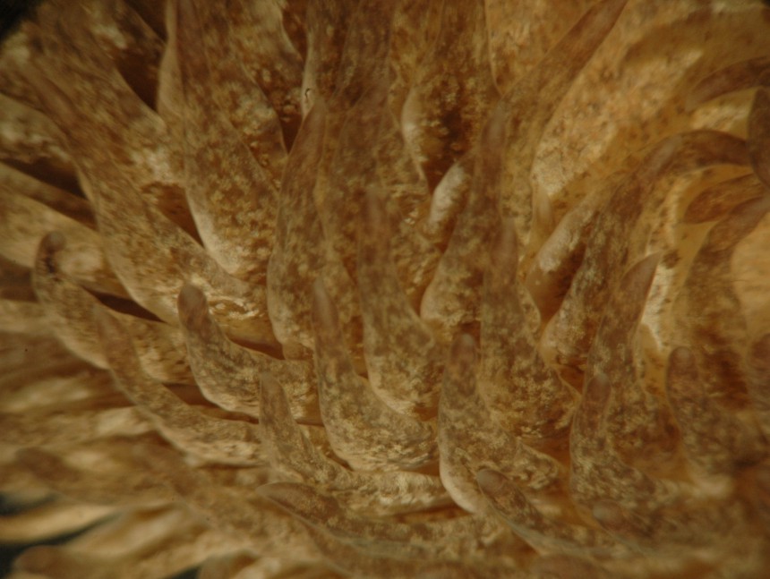
The cerata
are flattened, widest above the base, and taper to a point.
The hepatic
diverticula cannot be readily seen within them if there is pigment
present.
The tips often take on the coloration of their anemone food.
The
mid-dorsal band, which is cerata-free
but has light cororation on it, can be seen to the right.
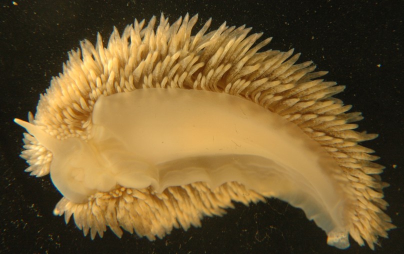
The foot tapers but is not drawn out into a long, sharp point.
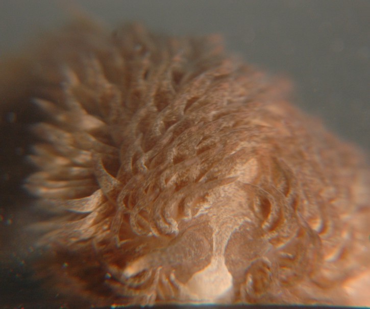
This head-on view shows the white triangle anterior to the rhinophores.
It also shows the smooth, tapering rhinophore
with a light-colored tip, and the fact that the rhinophore
seems to have a pore in the end. Notice also the cerata-free
band that runs mid-dorsally
and has light coloration (sometimes speckled or spotted).
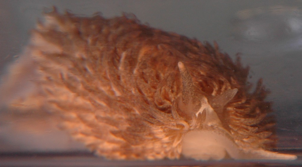
This head-on view shows the rhinophores
(the one on the right seems to have been injured and truncated), plus
one
of the two pedal
tentacles extends to the right.
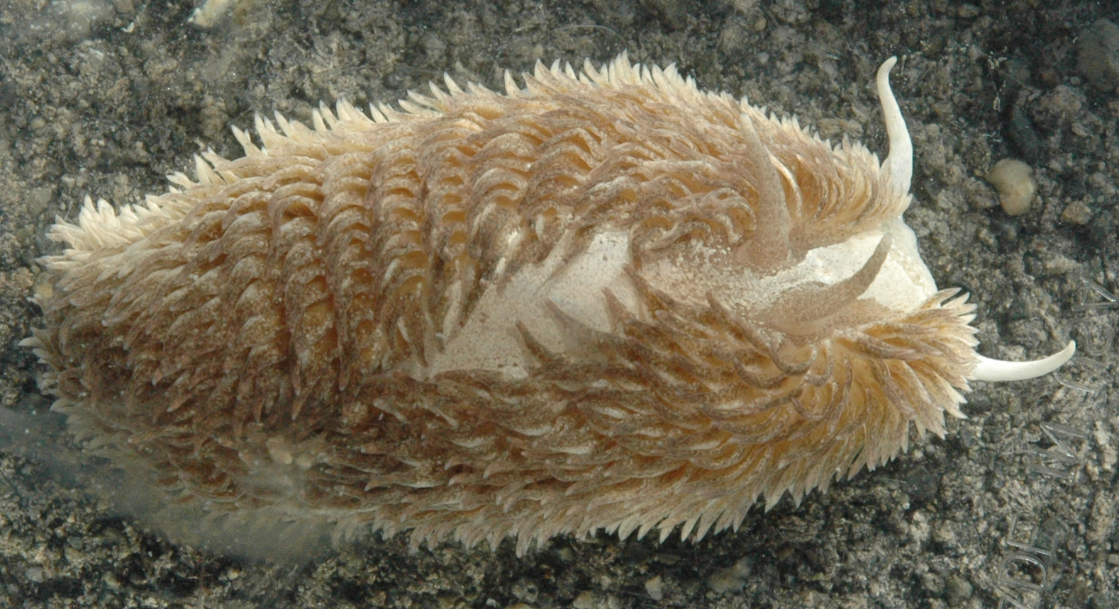
Another individual, 9 cm long including its oral tentacles, was found
on muddy intertidal sand at Padilla Bay June 2021 by Mason Fisher. The
warty, tapering, brown-spotted rhinophores plus the light-colored oral
tentacles are both plainly visible on the right, as well as the
triangular
cerata-free regions in front of the rhinophores and running down the
center
of the dorsum. Photo by Dave Cowles, June 2021
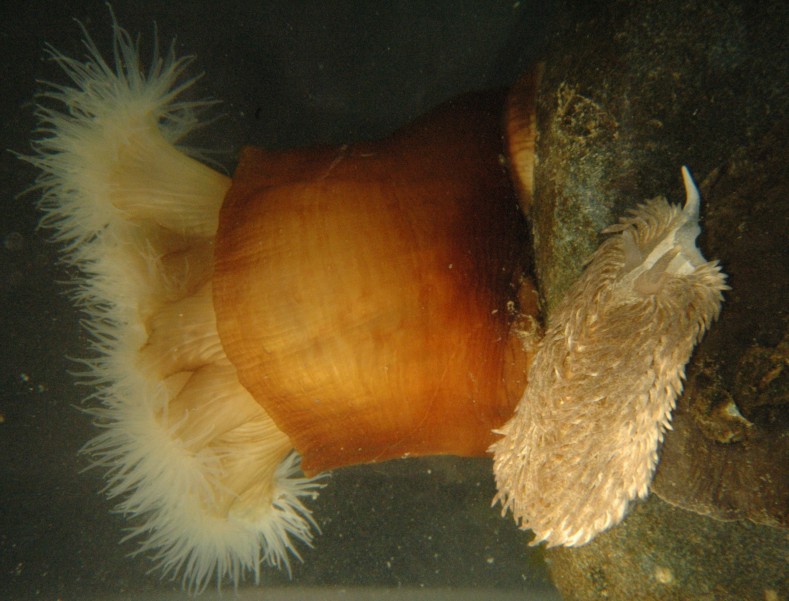
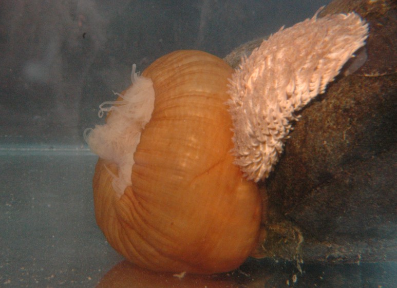
| In the sequence below, Aeolidia loui encounters and briefly attacks a Metridium giganteum anemone, eliciting the discharge of acontia from the anemone. Photos by Dave Cowles, 2008. | |
 |
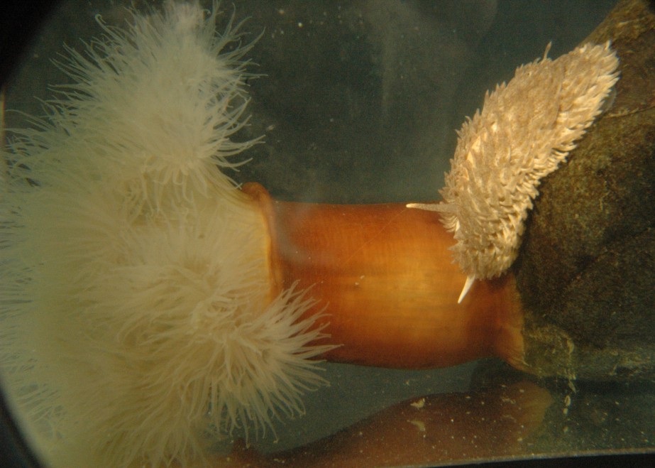 |
| The nudibranch's first encounter with the anemone | The nudibranch crawls out onto the column of the anemone. I did not see any copious quantities of mucus secreted, but that may be because I placed these two together. Earlier I had placed the nudibranch on the column of the anemone but it rolled up into a ball and dropped off. |
 |
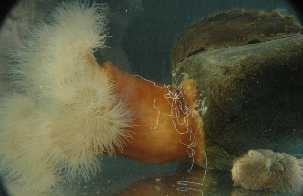 |
| Shortly after the nudibranch crawled out onto the anemone, the anemone rapidly contracted and closed up. The nudibranch turned away. | After the nudibranch left the anemone began opening up, then discharged multiple acontia from the column wall. |
Authors and Editors of Page:
Dave Cowles (2008): Created original page
CSS coding for page developed by Jonathan Cowles (2007)