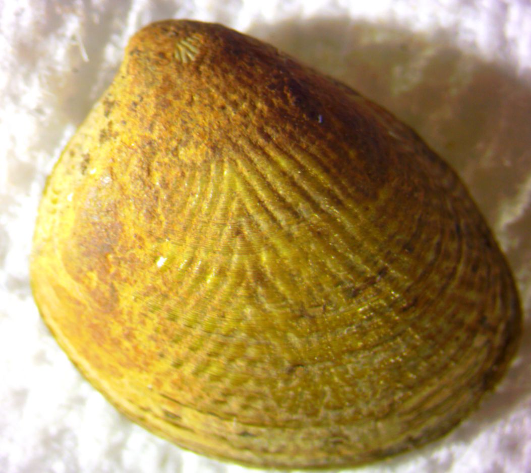
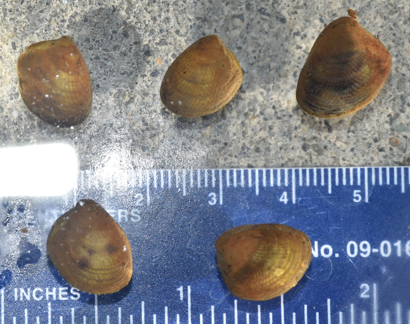
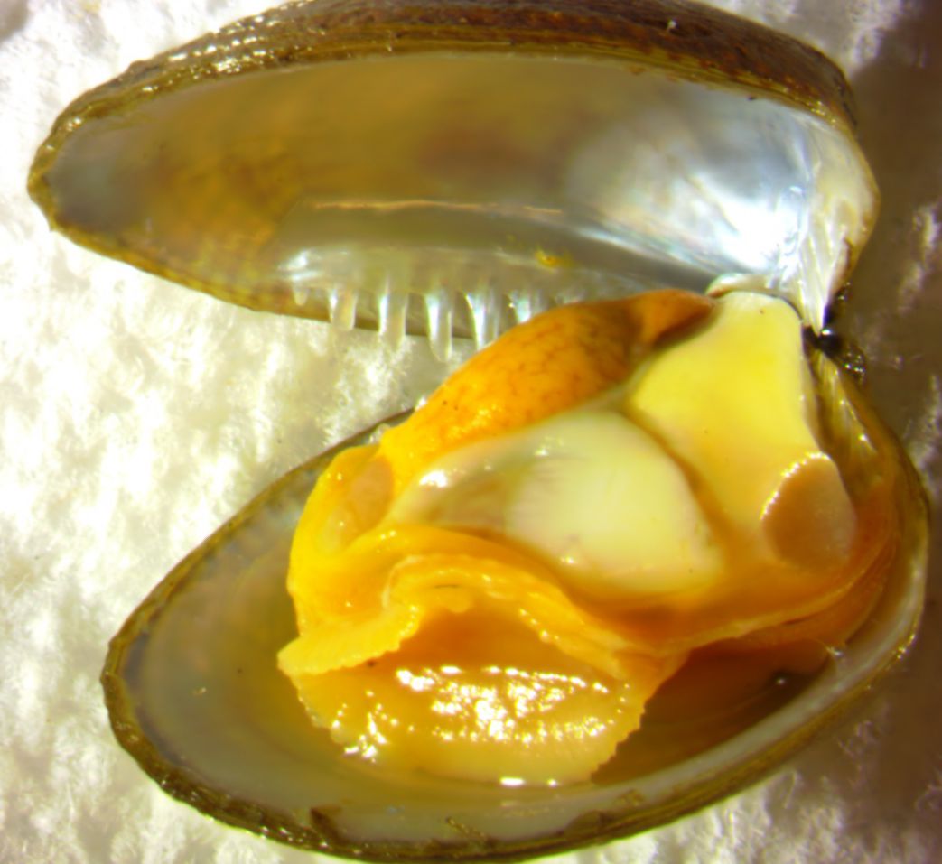
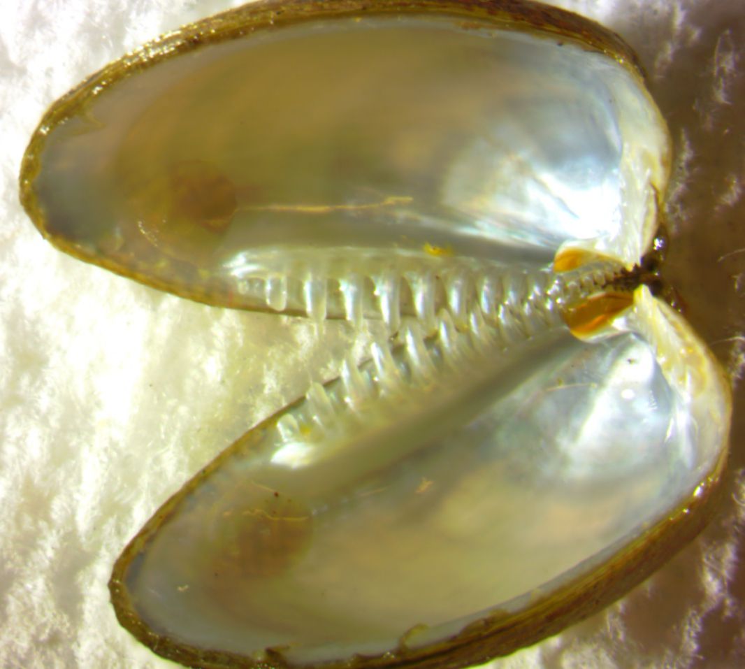
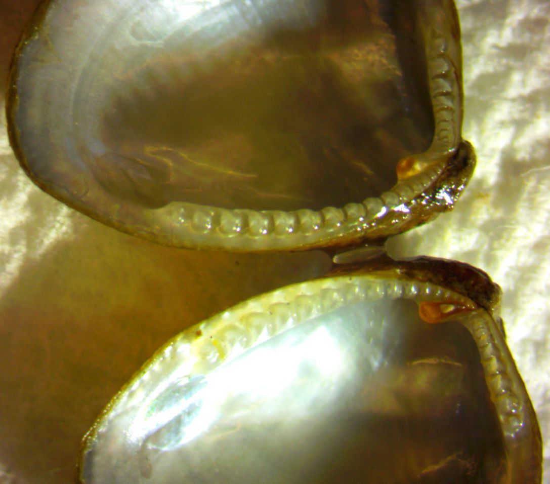
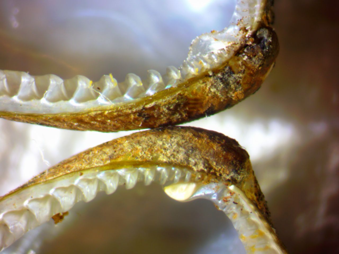
Description: Nut shells (Family Nuculidae) are small Protobranch clams with no siphons, an approximately oval shape with the anterior end somewhat squared off, and a pearly interior (photo). They have taxodont dentition on the shell (photo). Acila castrensis has a length approximately the same as the width, and both greater than the height. The outside of the valves are covered with a honey-colored, olive, or dark brown periostracum. The exterior sculpture of the shell is distinctive (see above): A series of radial ridges diverge ventrally from one another along the midline of the shell. Each ridge consists of a series of raised bumps, making a chevron-like pattern looking somewhat like a row of tents. Length to 2 cm, usually 15 mm or less. How to Distinguish from Similar Species: The small size and divaricate sculpture pattern on the shell is distinctive. Of clams with taxodont dentition, Ennucula tenuis is elongated and has no radial sculpturing. Geographical Range: East Pacific ocean, from Bering Sea, Alaska to Baja California, Mexico and in the Gulf of California. Depth Range: Subtidal, 5-400 m Habitat: Muddy-sand bottoms Biology/Natural History: Little is written about the biology of this clam. Protobranch clams do not filter feed with their gills as do other clams. Instead, they have much smaller, simple gills that are used mainly for respiration. For feeding, they extend two pairs of elongated labial palps to pick up edible particles from the sediment by ciliary action. The innermost pair (flanking the mouth) are called labial palps, while the two outermost palps are longer and called palp proboscides. It is the palp proboscides which are extended outside the shell, while the labial palps do later sorting of particles. Order Nuculida is mainly found in the deep sea. Stasek (1961) studied the complex ciliary patterns on the labial palps and concluded that the clams can capture food from the palp proboscides, from the labial palps themselves (via particles in the mantle cavity sticking to them), or from particles which stick to the gills and are passed on to the palps. Unlike most bivalves, the blood of this species contains hemocyanin (Morse, 1986, Mangum et al., 1987). The names of this clam refer to the distinctive chevron-like pattern of diverging ridges on the shell, which looks like an orderly camp of rows of tents.
References:Dichotomous Keys:Carlton, 2007 Kozloff, 1987, 1996 General References:
Scientific Articles:
Checa, Antonio G., Julyan H.E. Cartwright, and Marc-Georg Willinger, 2011. Mineral bridges in nacre. Journal of Structural Biology 176: pp 330-339 Kellogg, James L., 1915. Ciliary mechanisms of lamellibranchs with descriptions of anatomy. Journal of Morphology 26:4 pp 625-701. doi: 10.1002/jmor.1050260403 Mangum, C.P., J.L. Scott, K.I. Miller, K.E. Van Holde, and M.P. Morse, 1987. Bivalve hemocyanin: structural, functional, and phylogenetic relationships. The Biological Bulletin 173:11 pp 205-221. doi: 10.2307/1541873 Morse, M. Patricia, 1986. Hemocyanin respiratory pigment in bivalve mollusks. Science 231 pp 1302- Stasek, Charles R., 1961. The ciliation and function of the labial palps of Acila castrensis (Protobranchia, Nuculidae), with an evaluation of the role of the protobranch organs of feeding in the evolution of the bivalvia. Journal of Zoology 137:4 pp 511-538. doi: 10.1111/j.1469-7998.1961.tb06087.x Zardus, John D. and M. Patricia Morse, 1998. Embryogenesis, morphology and ultrastructure of the pericalymma larva of Acila castrensis (Bivalvia: Protobranchia: Nuculoida). Invertebrate Biology 177:3 pp. 221-244 Web sites: General Notes and Observations: Locations, abundances, unusual behaviors:
Authors and Editors
of Page:
Salish Sea Invertebrates web site provided courtesy of Walla
Walla University
|
|||||||||||||||