Proboscidactyla flavicirrata Brandt, 1835Common name(s): |
|
| Synonyms: | 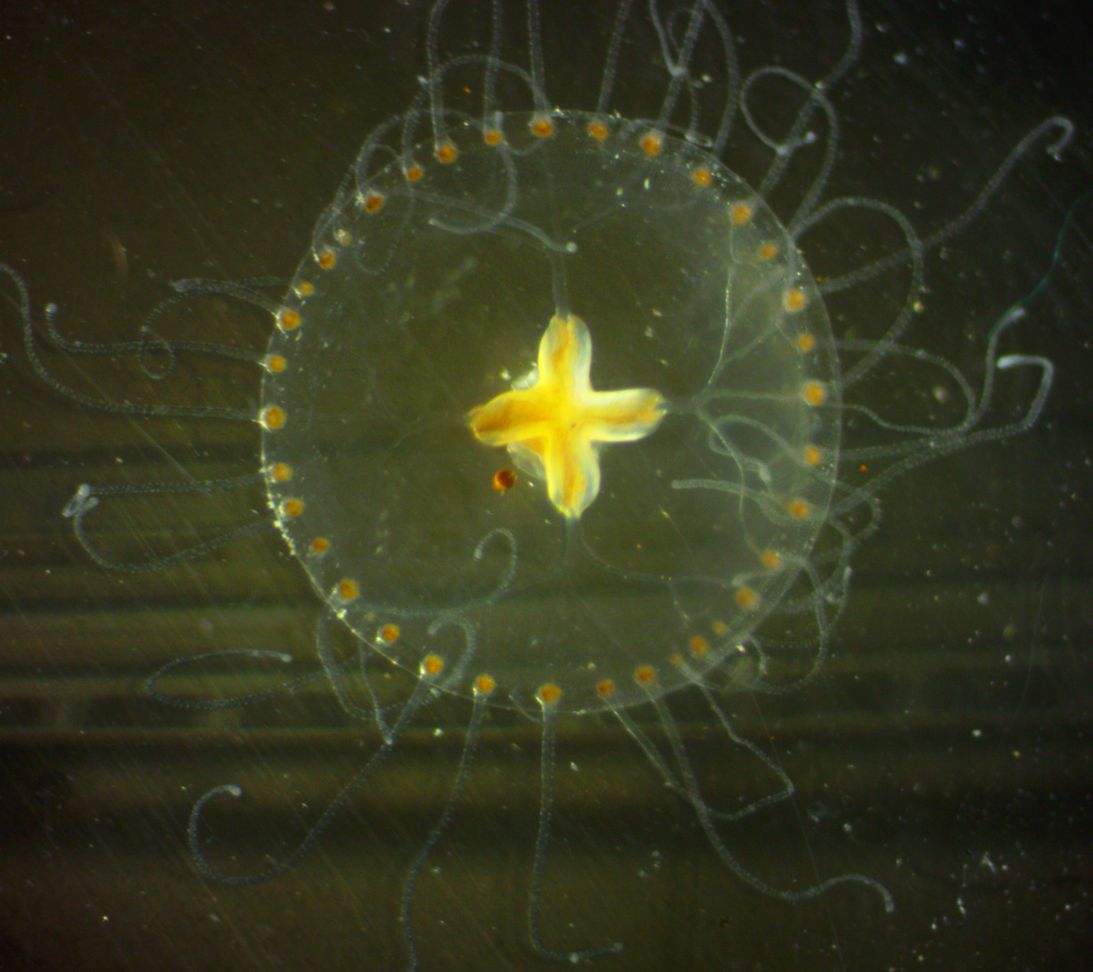 |
|
Phylum Cnidaria
Class Hydrozoa
Order
Hydromedusae
(Anthoathecatae, Anthomedusae)
Family Proboscidactylidae
|
|
| Proboscidactyla flavicirrata medusa, 3.5 mm in diameter, captured on a 6-m vertical plankton tow in the Strait of Juan de Fuca. A diatom is present on the bell. | |
| (Photo by: Dave Cowles, August 2014 ) | |
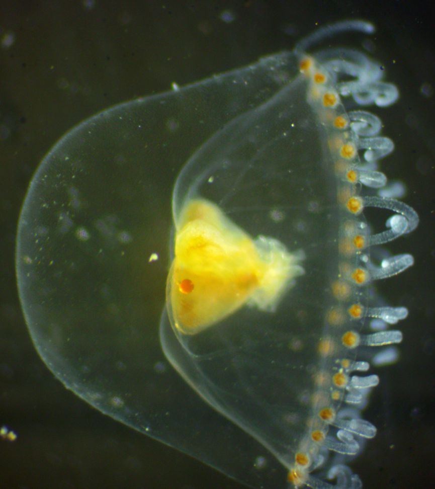
Description: This small hydromedusa has about 20-70 unbranched tentacles of similar size (40-72 when mature), which originate evenly around the margin of the bell. The tentacles have swollen tentacular bulbs at the base, and distinct knobs can be seen along the tentacles but the tentacles do not end in a thick cnidocyte knob (photo). The 4 radial canals branch dichotomously repeatedly and extend all the way to the bell margin (photo), so that by the time the ring canal is reached at the margin there are 50-70 radial canals. The jelly is thickest at the apex, and comprises about 1/2 of bell height (photo). The manubrium (photo) is not located on a long stalk, has strongly folded lips, and contains 4 paired gonads which cover it. Short clusters of cnidocytes exist on the exumbrella between the tentacles, on tracks and at varying distances slightly above the bell margin. Maximum bell height 10 mm. Diameter slightly greater than height, and the bell usually tapers rapidly from the margin (photo); but sometimes it is almost hemispherical.
How to Distinguish from Similar Species: Most other local hydroids do not have radial canals which branch dichotomously. Trichydra pudica has asymmetrically-branching radial canals.
Geographical Range: North Pacific from China and Japan to central Oregon. Rare in Oregon.
Depth Range: [Captured in a 60-m vertical plankton tow in the Strait of Juan de Fuca]
Habitat: Pelagic. Polyps are found on the tubes of Sabellid tubeworms (plumeworms)
Biology/Natural History: The medusa of this species has no statocysts.
The colonial polyp (hydroid) form of this species lives obligately as a stolon-based colony on the rim of Sabellid polychaete tubes such as Schizobranchia insignis ond Pseudopotamilla ocellata. The gastrozooid (feeding) polyps have only 2 long tentacles, which they extend toward the polychaete and steal food from its radioles. They also eat eggs that the polychaete releases. The polyps have a cnidocyte-studded, knoblike hypostome which is extended away from the polychaete tube. The knoblike hypostome combined with the 2 tentacles extending the other direction forms a shape much like the head and hands of a person praying or of a ballerina dancing. The nonfeeding polyps of the colony may be farther from the rim. The polyps are not enclosed in a hydranth and the gonophores are not protected by a gonotheca. The gonozooids have no mouth or tentacles. The medusae bud off and are released.
The planula
larva
of this species swims until it is drawn in by the feeding current
produced
by the cilia on the radioles
(plume) of the worm. When it contacts the radiole
it releases nematocysts which anchor it to the radiole;
then further attaches itself by mucus. When the worm later
withdraws
into the tube the planula
allows itself to be scraped off onto the rim of the tube, where it
attaches
and grows up into the adult hydroid
polyp colony. As the worm builds its tube longer,
the hydroid
extends its stolon
so that its feeding polyps
always remain at the mouth of the tube.
| Return to: | |||
| Main Page | Alphabetic Index | Systematic Index | Glossary |
References:
Dichotomous Keys:Carlton, 2007
Kozloff, 1987, 1996
General References:
Kozloff, 1993
Morris et al., 1980
Scientific Articles:
Mills, C.E., 1981. Diversity of swimming behaviors in
hydromedusae
as related to feeding and utilization of space. Marine
Biology 64:
pp 185-189
Web sites:
General Notes and Observations: Locations, abundances, unusual behaviors:
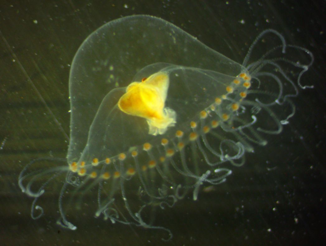
In this side view the swollen tentacular
bulbs at the base of the tentacles
and the knobs along their length, the short manubrium,
and the thick jelly which occupies about half the bell height can be
seen.
This view shows the dichotomously
branching radial
canals.
Authors and Editors of
Page:
Dave Cowles (2014): Created original page
CSS coding for page developed by Jonathan Cowles (2007)
Salish Sea Invertebrates web site provided courtesy of Walla
Walla University