Sylon hippolytes M. Sars, 1870Common name(s): |
|
| Synonyms: | 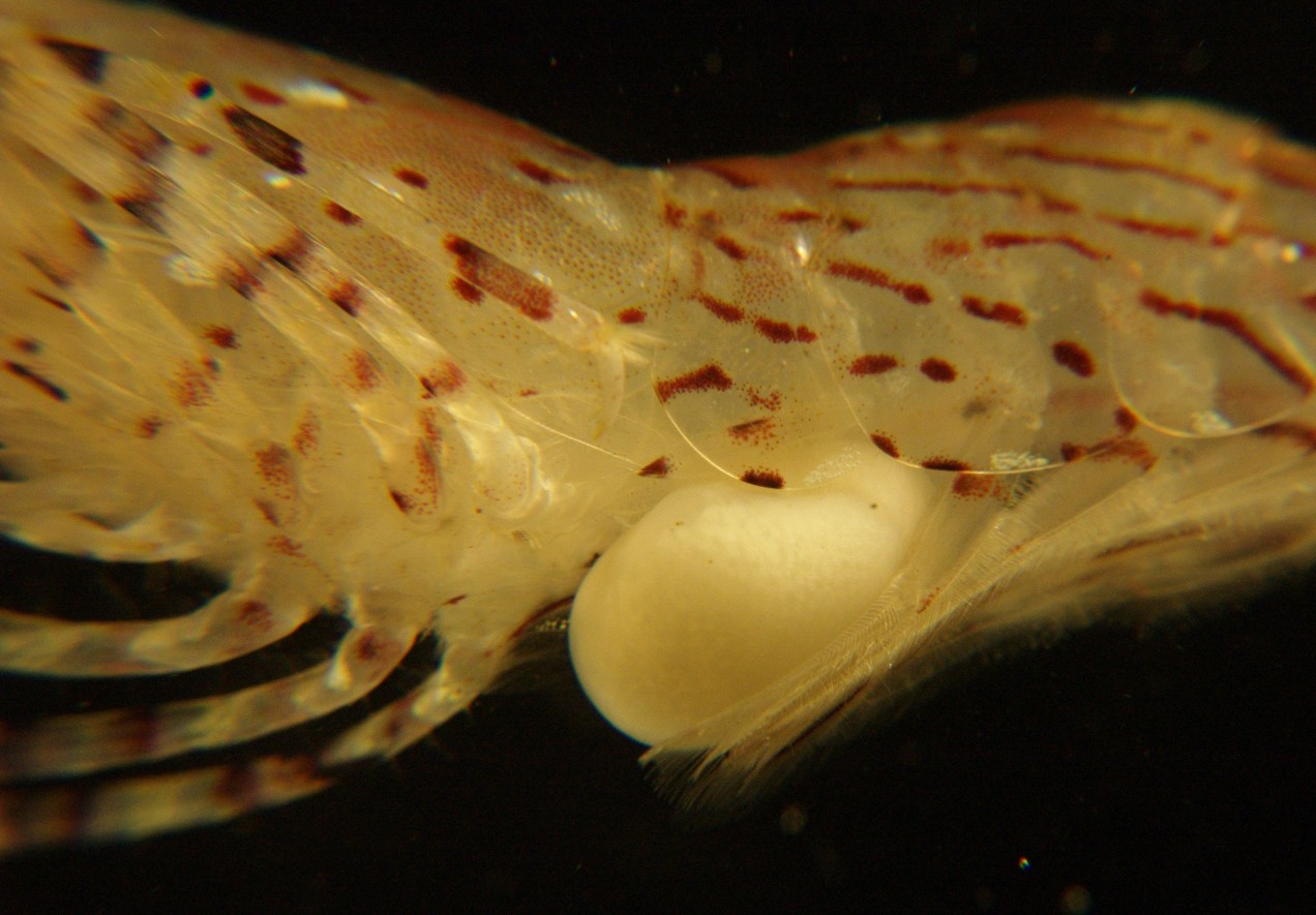 |
|
Phylum Arthropoda
Subphylum Crustacea
Class Maxillopoda
Subclass Thecostraca
Superorder
Rhizocephala
Order Akentrogonida
Family
Clistosaccidae
|
|
| Sylon hippolytes on a Pandalus goniurus shrimp captured at 75 m depth, San Juan Channel. | |
| (Photo by: Dave Cowles, August 2008 ) | |
How to Distinguish from Similar Species: This may be the only Sylon species. Its characteristic sack-like shape is distinctive.
Geographical Range: Arctic, Bering Sea, Chukchi Sea, North Pacific, Sea of Okhotsk, Sea of Japan, North Atlantic, Iceland, Greenland, Shetland Islands, Spitzbergen
Depth Range:
Habitat: Parasitic on shrimps from families Hippolytidae (Hippolyte, Heptacarpus, Spirontocaris, Caridion, Eualus, Lebbeus), Pandalidae (Pandalus), and Crangonidae (Sclerocrangon, Crangon, Metacrangon)..
Biology/Natural History: Rhizocephalan barnacles such as this species are bizarrely distorted parasitic barnacles. It was not even known that they were barnacles until the cypris larva in their life cycle was discovered. The eggs of Rhizocephalans usually hatch as a nauplius larva which metamorphoses into a cypris larva. Members of order Akentrogonida, however, such as Sylon hippolyte, apparently pass the nauplius stage in the egg and hatch as a cypris. The female cyprid settles onto a recently molted host or attaches to the host gill. She attaches to the host using a glue gland on her antennae. If she is a Kentrogonid, she then metamorphoses, losing her legs and eyes. She extrudes her tissue through the antenna or through her mouth through the host carapace into the host internal tissue. At that point she is called a "kentrogon". Akentrogonids such as Sylon do not form a kentrogon, but instead burrow directly into the host as a cypris larva. The injected barnacle enters the hemocoel (circulatory system) and grows into a ramifying rootlike structure called an "interna". The interna begins growing by sending out rootlike projections through the body of the host. These projections absorb nutrients from the host, and typically destroy the gonads (so they are called a parasitic castrator). The interna may grow very large and may actually become heavier than the host tissue. When she matures, a part of her body called the "externa" erupts through the exoskeleton of the host, usually on the ventral side of the abdomen near the gonads. The externa has a cavity for eggs and a place for males to attach. Male cyprids settle into the externa and metamorphose into a wormlike structure.
Unlike the family Sacculinidae (a Kentrogonid) in which the externa stops molting at maturity, Akentrogonid members of Family Clistosaccidae such as Sylon continue to molt the cuticle of the externa throughout life. That is one reason that the externa of Sylon almost always looks clean and un-fouled, as in the photo above, while the externa of sacculinids becomes fouled and dirty-looking (Hoeg, 1992).
Female shrimp which have this parasite species do not bear eggs, so the parasite is probably a parasitic castrator as are many Rhizocephalans.
| Return to: | |||
| Main Page | Alphabetic Index | Systematic Index | Glossary |
References:
Dichotomous Keys:Kozloff, 1987, 1996 (but not keyed)
General References:
Butler,
1980 (this is the main reference)
Lamb
and Hanby, 2005
O'Clair
and O'Clair, 1998
Scientific Articles:
Boschma, H., 1928. Rhizocephala of the north
Atlantic
region. Danish Ingolf Exp. 3(10). 49 pages.
Boschma, H., 1931. Papers from Dr. Th. Mortensen's
Pacific
Expedition 1914-1916. LV. Rhizocephala.
Viden.
Medd. Dan. Naturhist. Foren. Kobenhavn 89: 297-380
Calman, W.T., 1898. On a collection of Crustacea
from
Puget Sound. Ann. N.Y. Acad. Sci. II: 259-293
Hoeg, J. and J. Lutzen, 1985. Crustacea
Rhizocephala.--Marine
Invertebrates of Scandinavia 6: 1-92. Norwegian University
Press,
Oslo, Norway
Hoeg, Jens T., 1992. Chapter 6: Rhizocephala. pp 313-345 in
Frederick W. Harrison and Arthur G. Humes (editors), Microscopic
Anatomy of Invertebrates Volume 9: Crustacea. Wiley-Liss Publishers,
New York. ISBN 0-471-56116-9
Hoeg,
J. and A.V.
Rybakov, 1992. Revision of the Rhizocephala
Akentrogonida (Cirripedia),
with a list of all the species and a key to the identification of
familes.
Journal of Crustacean Biology 12(4) 600-609
Smith, G.W., 1906. Rhizocephala. Fauna
Flora Golfes
Neapel (Berlin) Monogr. 29. 123 pages.
Web sites:
General Notes and
Observations: Locations,
abundances, unusual behaviors:
I have not often seen this parasite.
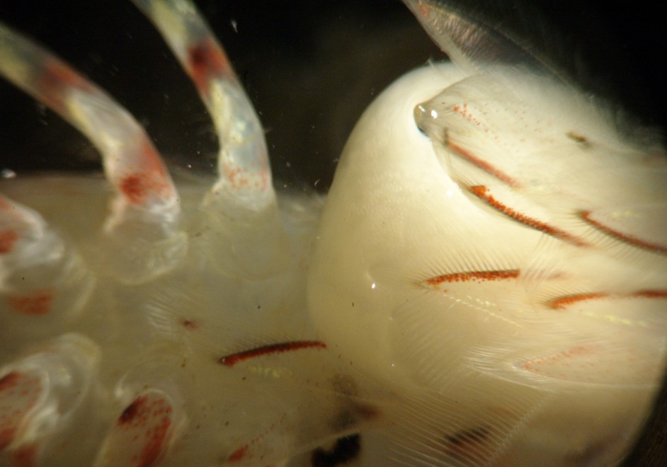
Ventral view of the parasite. The host's thorax is to the left. The parasite externa is erupting from the ventral side of the host's first abdominal segment. The second pair of host pleopods partly overlap the externa. The texture of the externa is smooth and clean but looks as if it may be packed with small eggs.
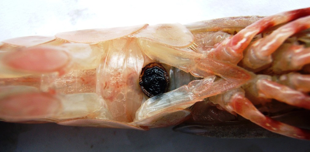
This externa scar on the abdomen of Pandalus platyceros
is the result of a Sylon
hippolytes parasitism. Photo provided by Hilary
Wood of the Alaska Dept. of Fish and Game.
Authors and Editors of Page:
Dave Cowles (2008): Created original page
CSS coding for page developed by Jonathan Cowles (2007)