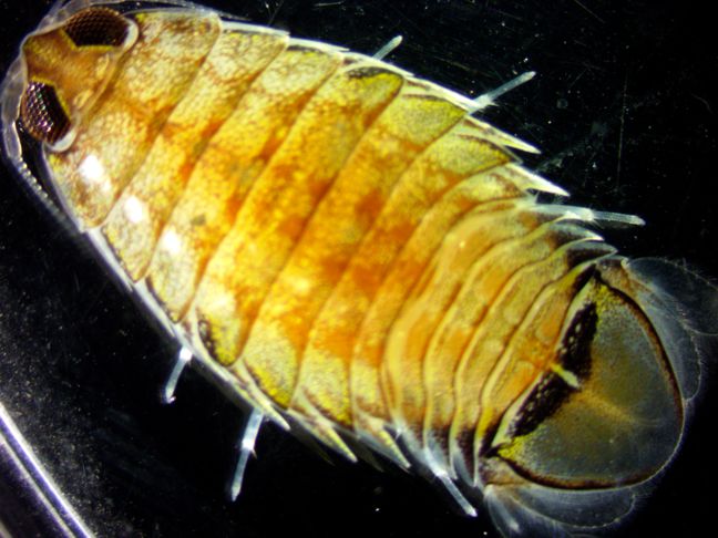
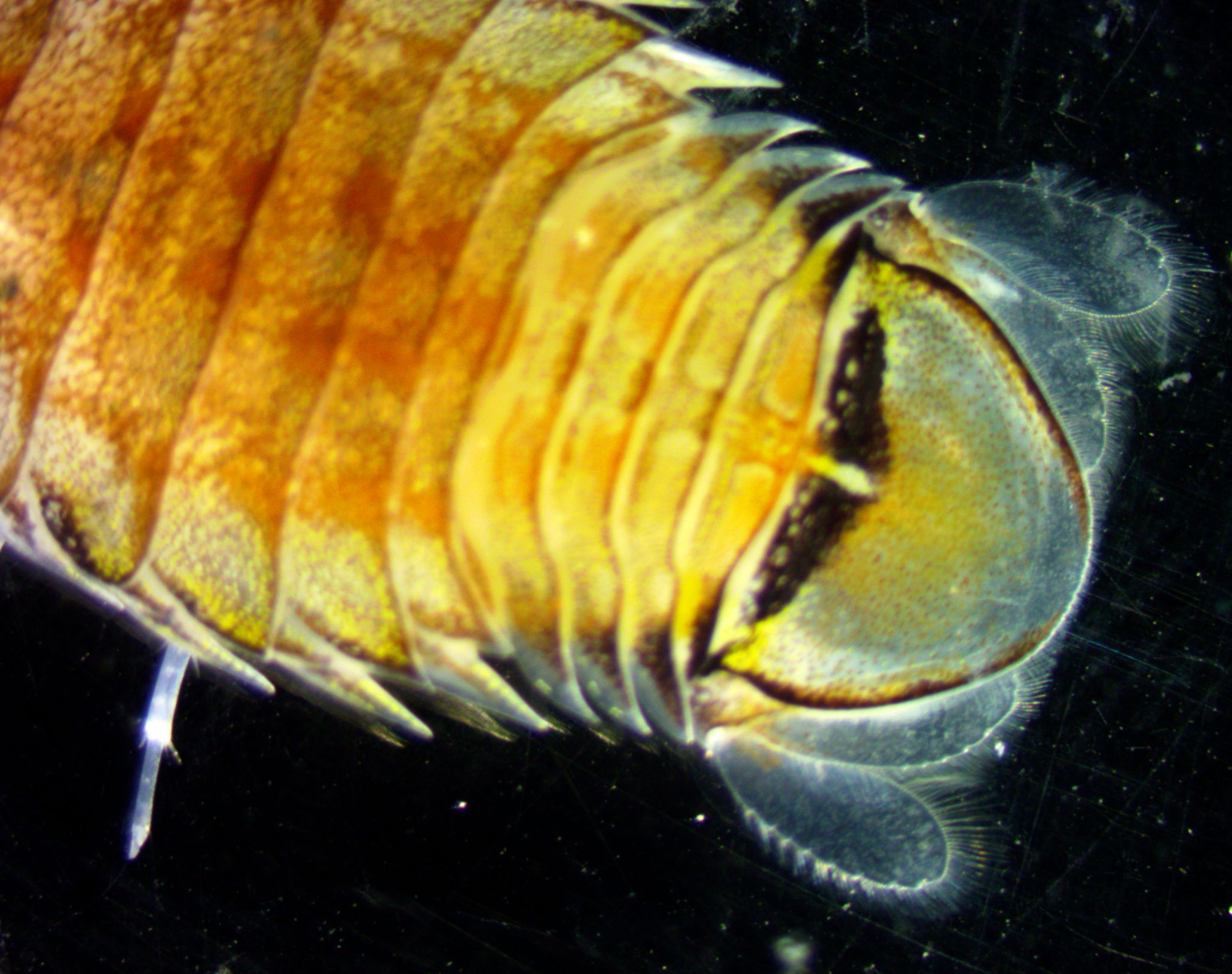
Description: As a member of suborder Flabellifera, this species has flattened uropods lateral to the pleotelson but not arching over it, and together with the pleotelson forming a caudal fan (photo). The body is not more than 5x as long as wide. This species is about twice as long as wide. Both rami of the uropods are well-developed and flattened and do not roll up into a ball (photo). They also do not extend past the posterior end of the pleotelson. The outer uropodramus is slightly shorter and narrower than the inner ramus. The outer margins of both rami are lined with long spines, and the inner margin with long setae. The species has 4 or 5 pleonites visible dorsally in front of the pleotelson (pleonite 1 is hidden beneath the last pereonite) (photo). The outer margins of most or all of the pereonites have points projecting posterolaterally. The dactyls of pereopods 1-3 are curved into hooks for holding on to the fish which they parasitize (photo). The propodus of these 3 pereopods has a large expansion on the medial side, with 6 teeth of about equal size (photo). The propodus and dactyl of these legs are approximately the same length. The last four pereopods have numerous spines (photo). The eyes are large and separated by a distance of approximately the diameter of the eye (photo). The flagellum of antenna 1 has only 4-6 articles and reaches approximately to the end of the peduncle of antenna 2 (photo). The flagellum of antenna 2 has 16 articles and reaches the posterior margin of the second pereonite. The rostrum is shaped like a truncated triangle; not drawn out into a spatulate shape with side projections (photo). The type specimen was reported to be brown with small black dots, although it was not described until several years after it was caught. Perhaps this golden color with white dots will change to brown with black dots upon preservation (see photo).
How to Distinguish from Similar Species: The very similar R. angustata has a propodus only about half as long as the dactyl on the anterior 3 grasping pereopods, and with only 4 instead of 6 teeth along the inner propodus margins. Rocinela tridens has a rostrum drawn out into a spatulate shape with side projections. R. belliceps has a pointed rostrum.
Geographical Range: East Pacific; as far north as Departure Bay, British Columbia but apparently not found as far south as California (not found in the Light and Smith manual). Thesingle type specimen (a male) was found in 1903 by the Albatross Expedition at station 4205 in Admiralty Inlet near Port Townsend, WA and described in by Harriett Richardson in 1905.
Depth Range: The type specimen was taken at 15-26 fathoms (27-48 meters). In 2024 we captured this species at 120 m depth in the San Juan Channel by otter trawl.
Habitat: Presumably near sandy or rocky bottoms or on bottom-dwelling fish
Biology/Natural History: This species parasitizes the gill regions of cartilaginous fish such as rays, and on bony fish such as halibut, rockfish, and other fishes. It may at times (such as this occasion) be found swimming freely in the water.
A related species, R. signata, has been known to repeatedly attack and bite human divers in the Caribbean. (Garzon-Ferreira, 1995. Bulletin of Marine Science 46:3 pp 813-815).
| Return to: | |||
| Main Page | Alphabetic Index | Systematic Index | Glossary |
References:
Dichotomous Keys:Kozloff, 1987, 1996
General References:
Scientific Articles:
Fee, A.R., 1920. The isopoda of Departure Bay and vicinity,
with
descriptions of new species, variations, and colour notes.
Contributions
to Canadian Biology and Fisheries 2.
Richardson, Harriet, 1905. Isopods from the Alaska Salmon Investigation. Bulletin of the Bureau of Fisheries Vol. XXIV, 1904 pp 209-221. (Original description is here)
Richaradson, Harriet, 1905. Monograph on the Isopods of North America. Bulletin of the United States National Museum No. 54, Washington Government Printing Office.
Web sites:
Schotte, M., B. F. Kensley, and S. Shilling. (1995 onwards). World
list of Marine, Freshwater and Terrestrial Crustacea Isopoda. National
Museum of Natural History Smithsonian Institution: Washington D.C.,
USA.
http://invertebrates.si.edu/isopod/
General Notes and Observations: Locations, abundances, unusual behaviors:
This dorsal
view of the pleotelson
shows the flattened uropods
lined with spines on the outer margin and setae
on the inner margin. It also shows the 4-5 pleonites
which are visible in front of the pleotelson.
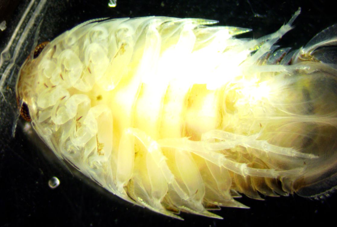
This view of the ventral side shows the spines on the posteriorpereopods.
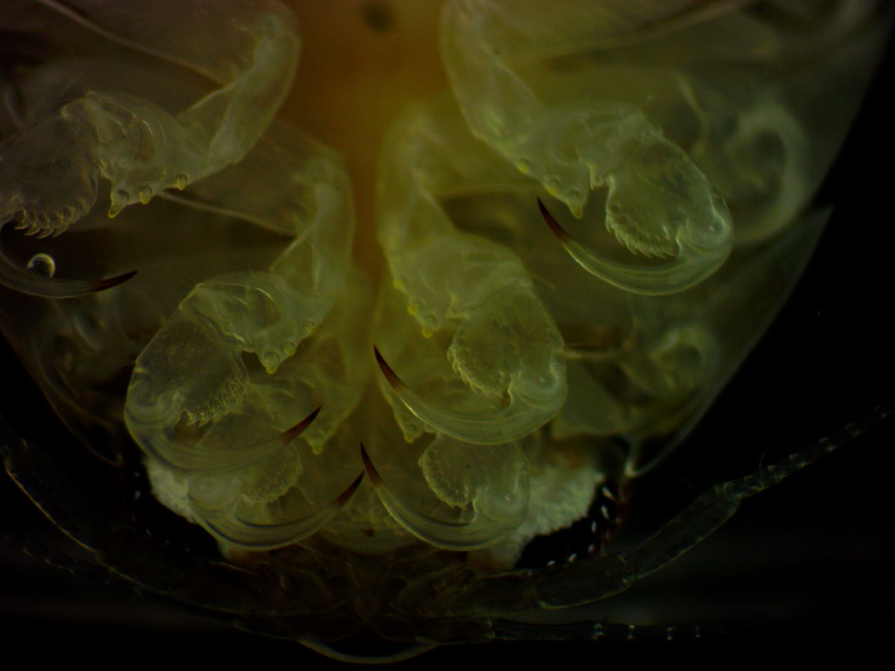
This ventral
view of the anterior
3 pereonites
show the curved dactyls,
which are about as long as the propodus.
It also shows the medial
expansions of the propodus
of these pereonites,
armed with 6 teeth. The merus
of these pereonites
has 3 blunt spines. Note also that the flagellum
of the first antenna has only 4-6 articles.
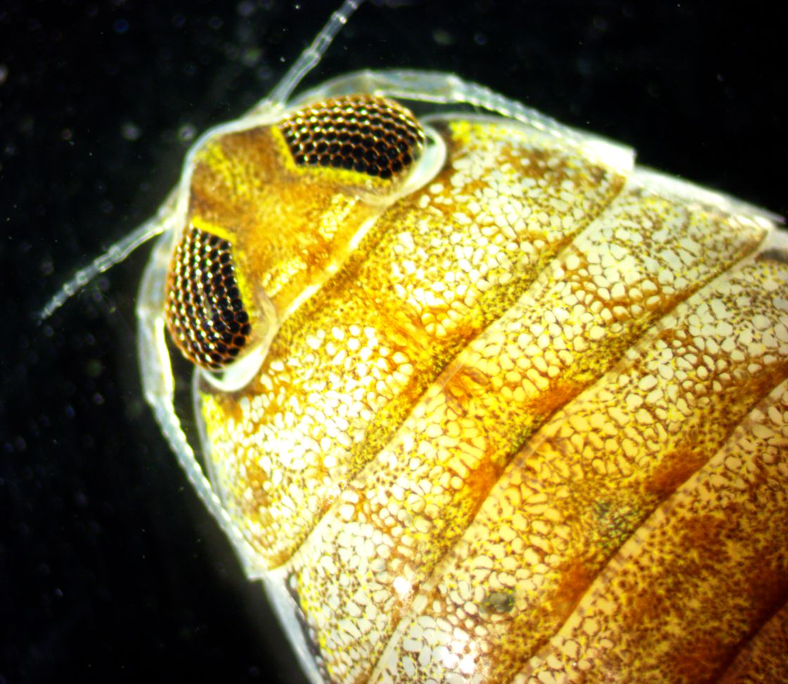
This dorsal view of the head shows the large eyes separated by about
the diameter of an eye; the truncated
triangular rostrum,
and the fact that the first antenna has only 4-6 articles
in its flagellum.
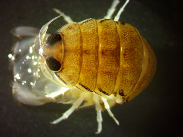
Here is the same individual several days after being preserved in
formalin.
The dark colors and black do indeed seem to be more prominent after
preservation.
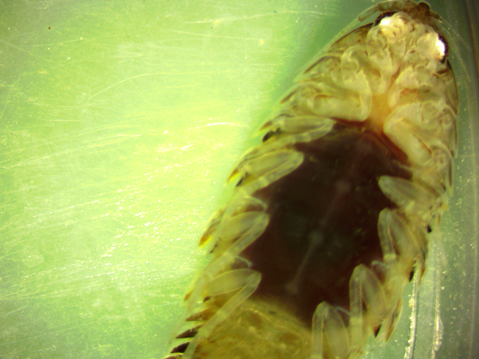
| The two photos below are a dorsal and ventral view of an egg-bearing female, bearing reddish eggs in her thoracic oostegites, captured by otter trawl at about 80-120 m depth in San Juan Channel July 2024. This video of the ventral side shows more details of the oostegites and the beating uropods (for breathing). |
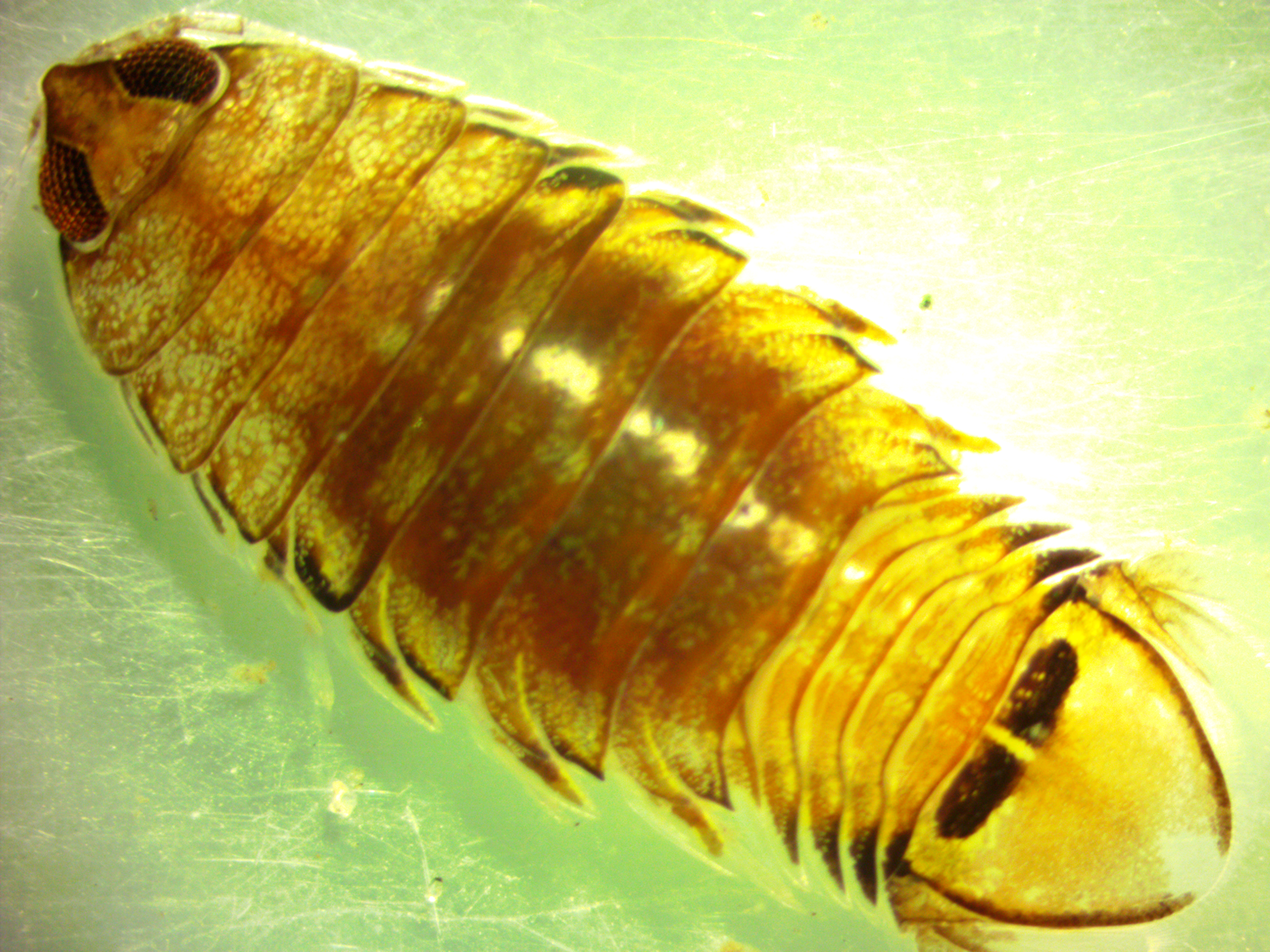 |
| Dorsal view above, ventral view with eggs below.
The eggs are carried
on the thorax, held in by flap-like oostegites which extend from the
base
of the legs.
Photos by Dave Cowles, July 2024 |
 |
