Metacrangon munita (Dana, 1852)Common name(s): Coastal spinyhead |
|
| Synonyms: Crago munita, Crangon munita | 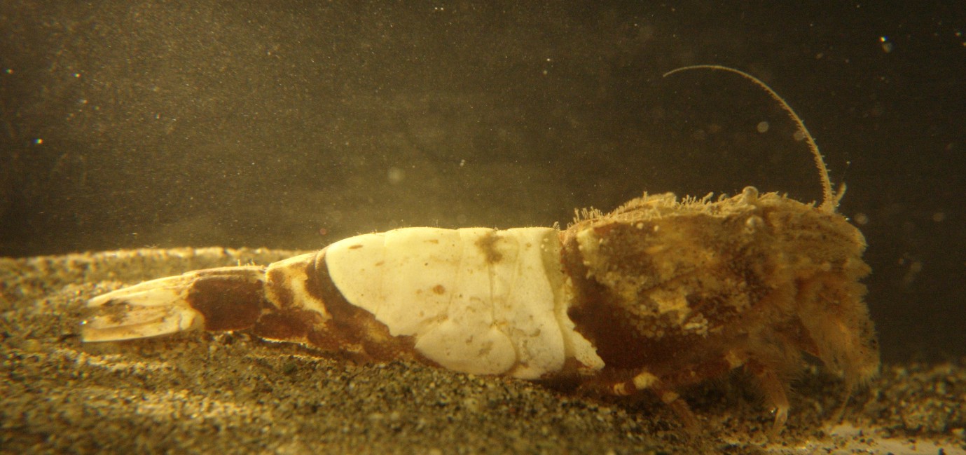 |
|
Phylum Arthropoda
Subphylum Crustacea
Class Malacostraca
Subclass Eumalacostraca
Superorder Eucarida
Order Decapoda
|
|
| Metacrangon munita, about 4 cm long, captured at 75 m depth in the San Juan Channel | |
| (Photo by Dave Cowles, July 2008 ) | |
How to Distinguish from Similar Species: The coloration of this species is distinctive. Metacrangon variabilis has a prominent longitudinal mid-dorsal ridge on abdominal segments 3-5 and the spine of its antennal scale is longer than the lamella. The anterior median carapace spine in Metacrangon acclivis is larger than the posterior spine and extends beyond the bases of the eye orbits. The pleura 1-3 of Metacrangon spinosissima have ventrally directed spines. Mesocrangon munitella has two submedian dorsal spines, and other genera of crangonids usually have none.
Geographical Range: Central Alaska to southern CA
Depth Range: 12-230 m
Habitat: Sand bottoms
Biology/Natural History: This species is nocturnal. Sometimes parasitized in the gills by the isopod Argeia pugettensis.
Morphologically (not by color) this species is similar to Metacrangon richardsoni from New Zealand.
Genus Metacrangon
have
two median and one submedian spine on the carapace.
The gastric region of the carapace
is depressed below the other regions. The term "spinyhead"
refers
to the median and submedian carapace
spines (and other spines as well). Most of the spines are on
the
end of a supporting carina,
or ridge.
| Return to: | |||
| Main Page | Alphabetic Index | Systematic Index | Glossary |
References:
Dichotomous Keys:Kozloff, 1987, 1996
Wicksten, 2009
General References:
Butler,
1980
Jensen,
1995
Lamb
and Hanby, 2005
Scientific Articles:
Web sites:
General Notes and Observations: Locations, abundances, unusual behaviors:
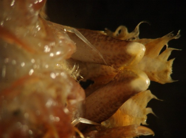
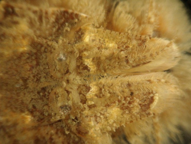
This ventral view of the head shows that the first pair of pereopods,
held closely below the head in this view, is subchelate.
In this side view of the carapace, the short rounded rostrum and two median dorsal spines can be seen. The dorsal profile of the carapace is highest at the posterior median spine and descends anteriorly all the way to the rostrum in a female, while in the male the dorsal profile remains high until the anterior median spine, then descends to the rostrum. From this view and the animal's size I conclude that this is a female. Also note that pereopod 5 (the last walking leg) does not have a broad and flattened dactyl. On the side of the carapace can be seen the light-brown submedian spine, and a hepatic spine just below and forward of it.
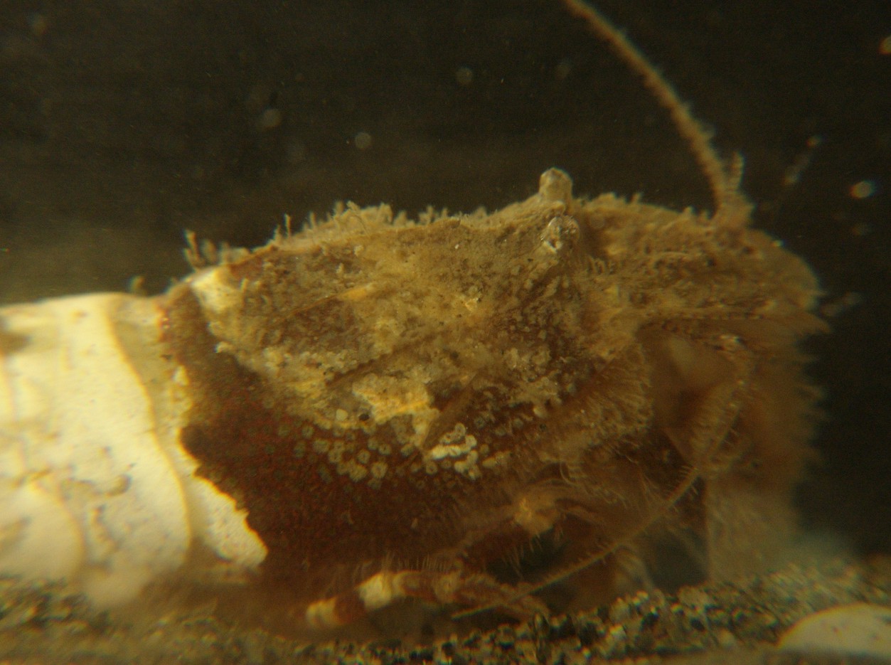
The many spines and setae and the disruptive coloration of the carapace of this species makes it hard to see structures up close. This is a dorsal view of the carapace, with the head facing right. The two eyes can be seen on the right. Between them is the small rostrum, then the mid-dorsal ridge runs back to the left. The first mid-dorsal spine is visible in the light area at the left of the picture. In front of the mid-dorsal spine and directly behind the eyes on each side is a single sub-median spine on each side, colored light brown and just to the left of center in this view.
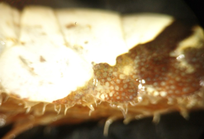
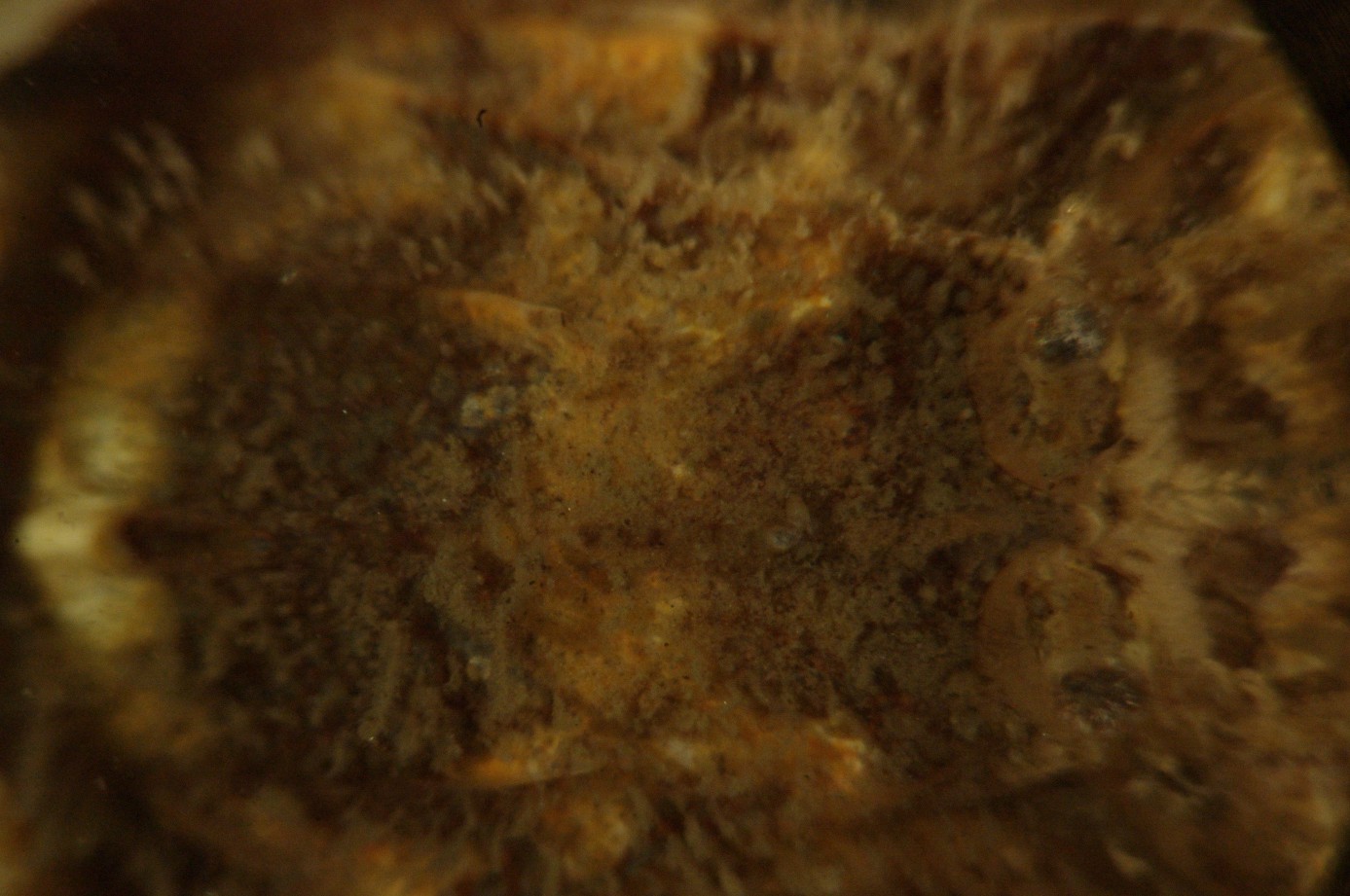
This side view of abdominal segments 2-4 shows that there are no ventrally-directed spines on segments 1-3 and no prominant longitudinal mid-dorsal ridge on segments 3-5. The first and third pleura have a depression that the larger 2nd pleuron fits over.
This dorsal view of the head shows the antennal
scale (2nd antenna). Note that the lamella
of the scale extends past the antennal
spine.
Authors and Editors of Page:
Dave Cowles (2008): Created original page
CSS coding for page developed by Jonathan Cowles (2007)