Ammothea verenae Child, 1987Common name(s): |
|
| Synonyms: Scipiolus thermophilus Turpaeva, 1988 | 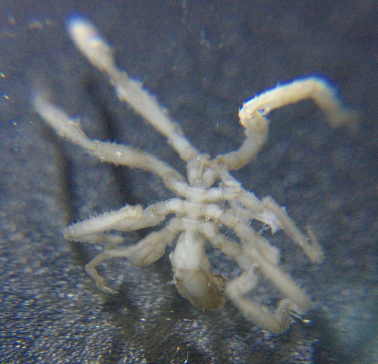 |
|
Phylum Arthropoda
Subphylum Chelicerata
Order Pantopoda
Family Ammotheidae
|
|
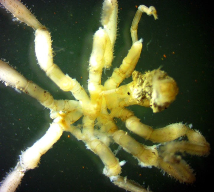
| A female Ammothea verenae pycnogonid, about 1 cm long from tip of proboscis to tip of abdomen. Collected by Kirt Onthank from Endeavor hydrothermal vents on Juan de Fuca Ridge off Washington. The large proboscis is at the bottom of the photo and the small abdomen is at the top. Alcohol-preserved specimen. | |
| (Photo by: Dave Cowles, Feb 2014) | |
Description: Order Pantopoda is the only Order in Class Pycnogonida. Family Ammotheidae is one of the largest and most variable families of pycnogonids. Members of Family Ammotheidae usually have chelifores (anterior appendages which arise from the cephalon and usually bear chelae), but some species have no chelifores or have no chelae on them. The palps have 1 to 10 segments. Both males and females in this family have 9 to 10-segmented ovigers, which are larger in males. This species has a slender trunk, the segments of which flare out into a cowling posteriorly (photo). The abdomen is long (for pycnogonids), and curves downward (photo). The lateral processes (serve as the bases for the legs) from the trunk segments are separated by less than half their diameters. The legs are long and have many setae (photo). Males have dense clusters of setae on their legs, while setae on the legs of females are much sparser (photo). The third coxae also have long ventral setae. The distal segments on different legs are similar to one another: The tarsus is very short (photo); the propodus is slender and curved (photo) (photo). The main claw at the end of the legs (photo) has several long auxiliary claws. The ocular tubercle is a low cone at about the middle of the neck (narrowed posterior part of cephalon), without eyes (photo). The species has a long proboscis with constrictions near the middle and at the end (photo). The chelifores on this species are very short and without chelae (photo). The palpi (photo) have 9 segments, the last 5 of which have many ventral setae. Oviger segments 4 and 5 are nearly equal in length. Strigilis segments 6-8 have many long lateral setae and segments 8-10 have 1-2 small denticulate spines on the end. The ovigers of females are smaller than those of males. The eggs (carried on the ovigers of the male) are only about 1/4 the diameter of the oviger segments they are attached to, and are carried in large clusters (photo). The cement gland (which is located on the dorsal surface of of the femur of the legs of males) opens as a tiny pore on the dorsodistal end of the femur.
How to Distinguish from Similar Species: This species is quite different from other members of the genus by being blind, by having no dorsomedian tubercles on the trunk segment cowls, and the propodus is similar on the anterior and posterior legs. Another common genus of pycnogonid found at hydrothermal vents isSericosura. Local shallow-water species of Ammothea have no strigilis.
Geographical Range: Northeastern Pacific: Juan de Fuca Ridge around Axial Seamount, Endeavor Segment, and Explorer Ridge
Depth Range: Around 2000 m
Habitat: Near hydrothermal vents
Biology/Natural History: Common near hydrothermal vents on the Juan de Fuca Ridge. Often encrusted with metal sulfides.
Segmentation in the legs of pycnogonids: The base is
connected
to a lateral process of the trunk. The first 3 segments
(usually
short) are coxae
1-3,
then a longer femur,
then long tibia
1 and
2, tarsus
(short),
propodus,
claw (which has a larger principal claw and may have paired, usually
smaller
auxiliary claws projecting from the same base)
| Return to: | |||
| Main Page | Alphabetic Index | Systematic Index | Glossary |
References:
Dichotomous Keys:General References:
Scientific Articles:
Child, C.A., 1987. Ammothea-verenae and Serocosura-venticola, 2 new hydrothermal vent-associated pycnogonids from the northeast Pacific. Proceedings of the Biological Society of Washington 100:4 pp 892-896
Web sites:
General Notes and Observations: Locations, abundances, unusual behaviors:
My thanks to Kirt Onthank for providing me with the preserved specimens shown on this page.
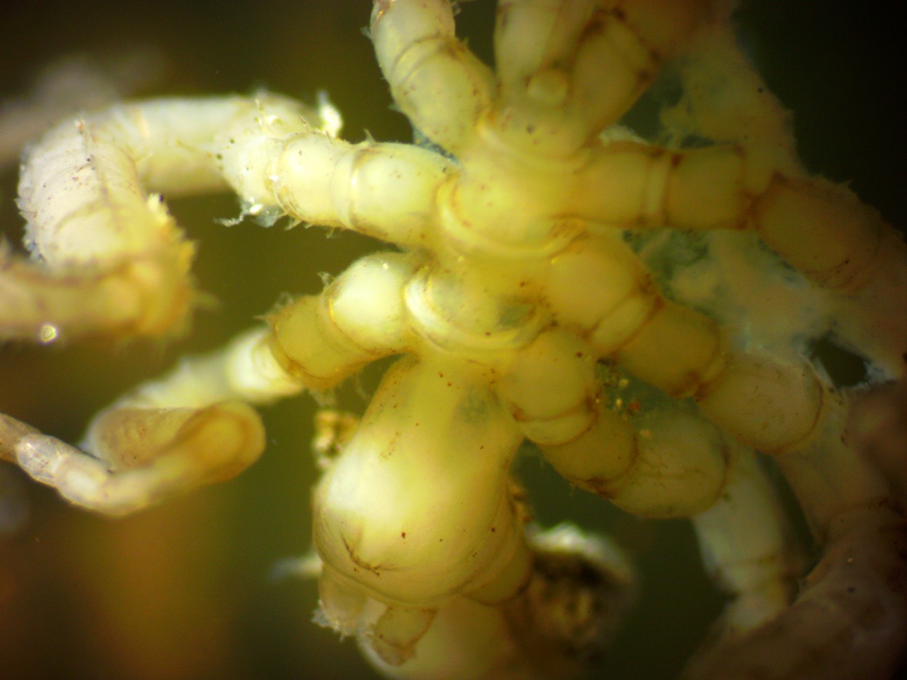
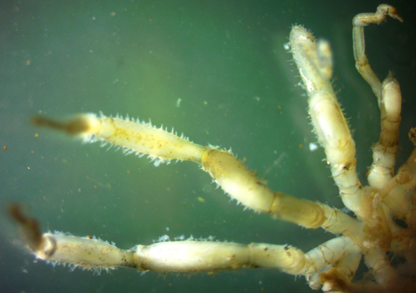
This dorsal view of the trunk and base of the head (cephalon) of this female shows several features. The animal is facing the bottom of the photo. Notice how the posterior ends of the trunk segments flare out. Other members of this genus also have dorsal tubercles arising from this flare but this species does not. Each trunk segment has lateral extensions to which the legs are attached. The three short coxae (basal segments) of most legs can be seen, then a longer femur which often extends upward while walking. The first and second tibia usually extend downward from the end of the femur so the animal walks with legs curved out to the sides similar to spiders. The ocular tubercle can be seen dorsally on the neck (base of the cephalon) near the bottom of the photo but note that it has no eyes. The stubby chelifores with undeveloped chelae can be seen just below the ocular tubercle. The base of the left palp can be seen exiting the cephalon to the right of the chelifores.
This dorsal view of the rear portion of the same animal (female) shows the abdomen, which is larger than seen on most pycnogonids, arising dorsally on the last thoracic segment and curving down between the hind legs.
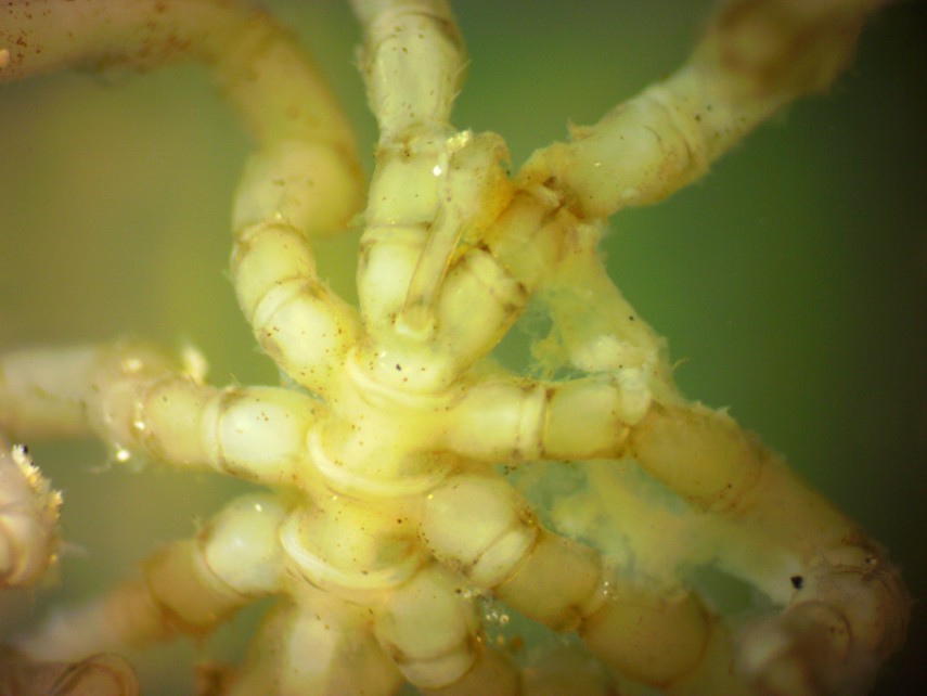
This dorsal view of a male shows similar features. This male is carrying a large mass of egg clusters ventrally to the trunk. Anterior is to the right.
| The photos below allow a comparison of the setation on male vs female legs | |
 |
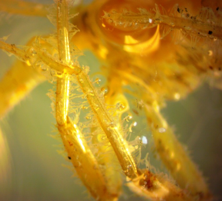 |
| The setae on female legs are more sparse and especially along the edges. | The legs of males have much more setae. |
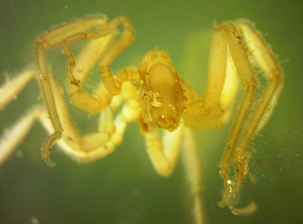
This view of the front of a female shows the constriction near the middle and at the end of the proboscis, the triangular mouth opening at the end of the proboscis bordered by 3 teeth, the palps along each side of the proboscis, and the stubby chelifores dorsally on the cephalon behind (which look almost like eyes in this view). Several pieces of debris are clinging to setae on the palps. Notice on the second right leg (visible on the left of the photo) one can clearly see the claw at the end, then the curved propodus, short tarsus, long ascending tibia 2 and 1, descending femur, and coxae 3 and 2. Coxa 1 is hidden behind the first leg.
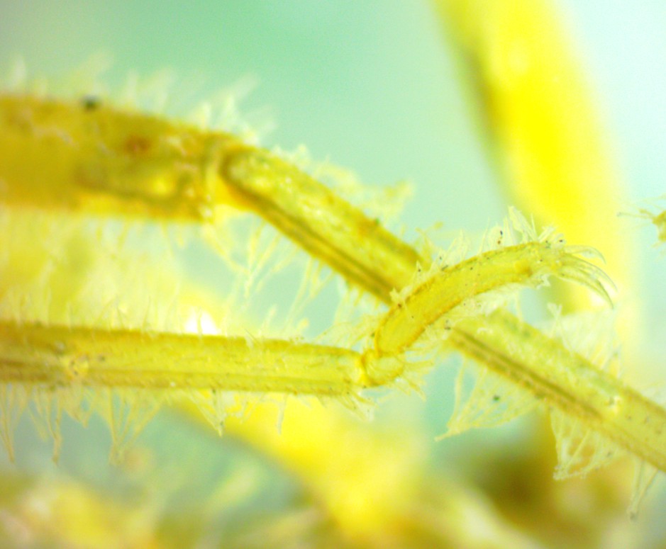
This photo shows the curved propodus and claw of a male.
This view of the underside of a female shows the ovigers
attached to the underside of the neck (rear of the cephalon)
and carried under her body. The proboscis
(to the right) has debris adhered to the ventral side. Note
the curved
strigilis
at the end of the ovigers.
The strigilis
is
used for grooming. The oviger
on males is larger and has more setae
on the strigilis.
There are also more setae
on the legs of males.
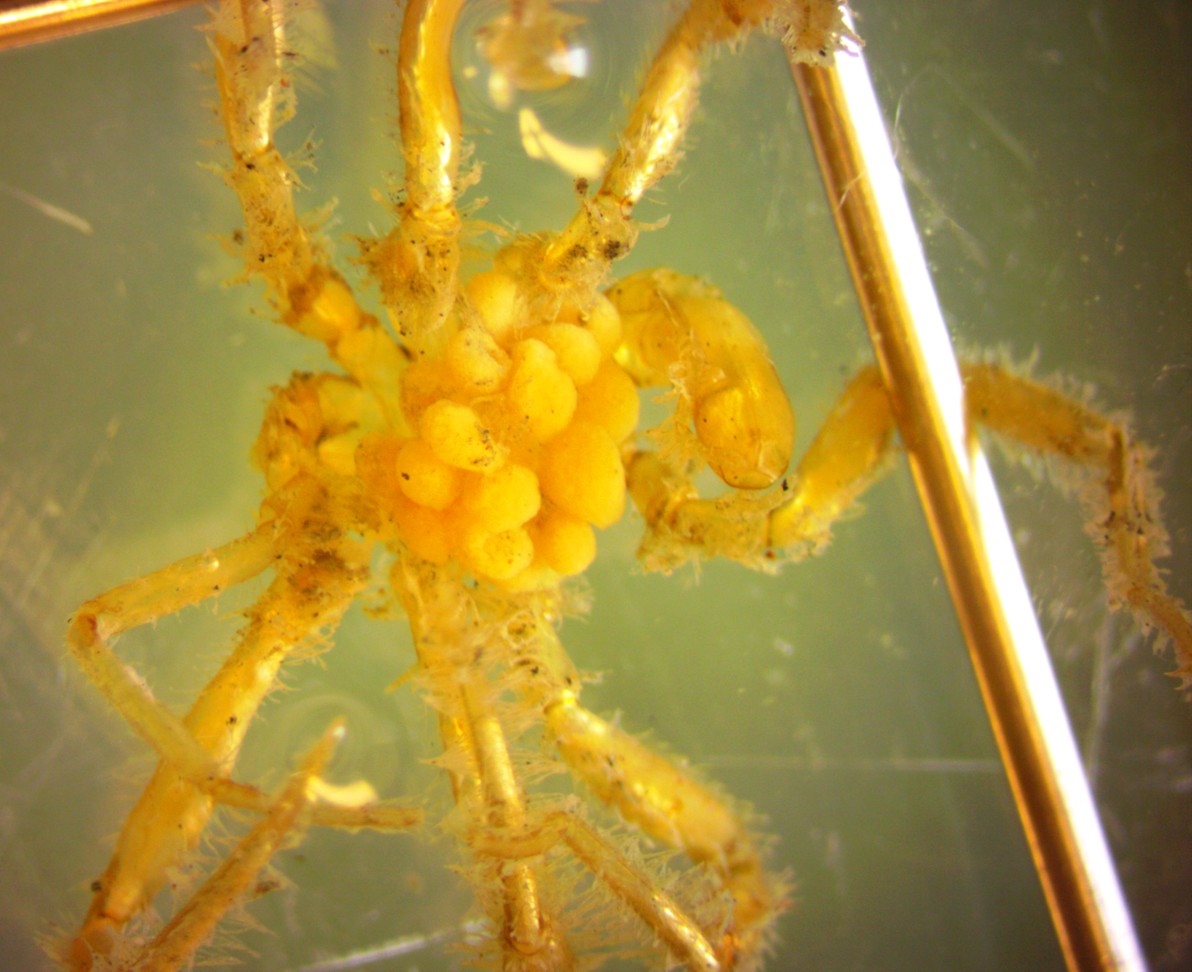
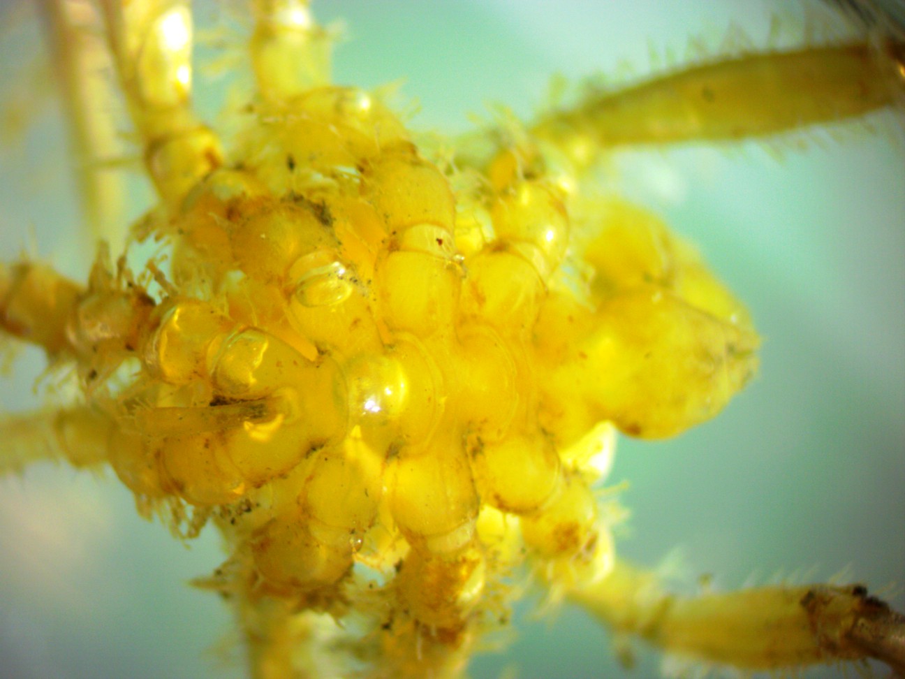
This
ventral view of a male (preserved in alcohol) shows he is carrying a
large cluster of eggs. Presumably they are attached to the
ovigers but they are so large the ovigers are completely obscured.
The
description of this species in Desbruyeres et al. says the eggs are
small--only about 1/4 the diameter of the oviger segments, but are
carried in large round clusters. The clusters can clearly be
seen
here.
Authors and Editors of Page:
Dave Cowles (2014): Created original page
CSS coding for page developed by Jonathan Cowles (2007)
Salish Sea Invertebrates web site provided courtesy of Walla
Walla University