Gattyana ciliata Moore, 1902Common name(s): Ciliated scaleworm |
|
| Synonyms: | 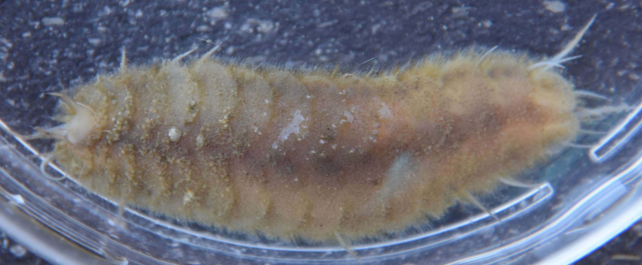 |
|
Phylum Annelida
Order Phyllodocida
Superfamily
Aphroditacea
|
|
| Gattyana ciliata, 5 cm long, captured by otter trawl at 80 m depth in San Juan Channel. Head is to the left, and the right front scale (elytra) has detached. Note the fuzzy appearance on the scales, which is due both to the texture of the scales themselves and to debris and diatoms which seems to attach to them. | |
| (Photo by: Dave Cowles, July 2023) | |
Description: Polynoids (scaleworms) are a large family of actively-foraging or symbiotic polychaetes with much or all of their dorsal side covered with scale-like elytra. Members of genus Gattyana have 15 pairs of elytra and 35-40 segments, a bilobed anterior edge of the prostomium, plus their lateral prostomial antennae are inserted directly ventrally to the median antennae. Gattyana ciliata has 15 pairs of elytra which cover the entire dorsal side (see photo above), but less than 50 segments as visible on the ventral side (photo). The neurosetae are thick, simple, and amber (photo), and none of the neurosetae are bifid at the tip. The notosetae are capillary setae, much narrower than the neurosetae (photo). The lateral antennae of the prostomium are inserted ventrally to the medial antennae rather than at the anterior edge of the prostomium (photo). The exposed dorsal surface of the elytra do not have distinct, polygonal subsections but they do have several fairly large conical tubercles with truncated summits, especially along the free posterior margin of the elytra (photo). Other parts of the elytra have various smaller bumps and projections. Length to 8 cm.
How to Distinguish from Similar Species:Gattyana cirrosa and several other species has notosetae of two types, with the most dorsal thick and blunt, plus the elytra do not have the large conical tubercles along the posterior edge.
Geographical Range: Alaska to Washington on the NE Pacific coast. Also reported from Japan.
Depth Range: Subtidal
Habitat: Travels over a variety of habitats, including soft and rocky substrates.
Biology/Natural
History:
| Return to: | |||
| Main Page | Alphabetic Index | Systematic Index | Glossary |
References:
Dichotomous Keys:Kozloff, 1987, 1996
General References:
Lamb
and Hanby, 2005
Scientific Articles:
Web sites:
General Notes and
Observations: Locations, abundances,
unusual behaviors:
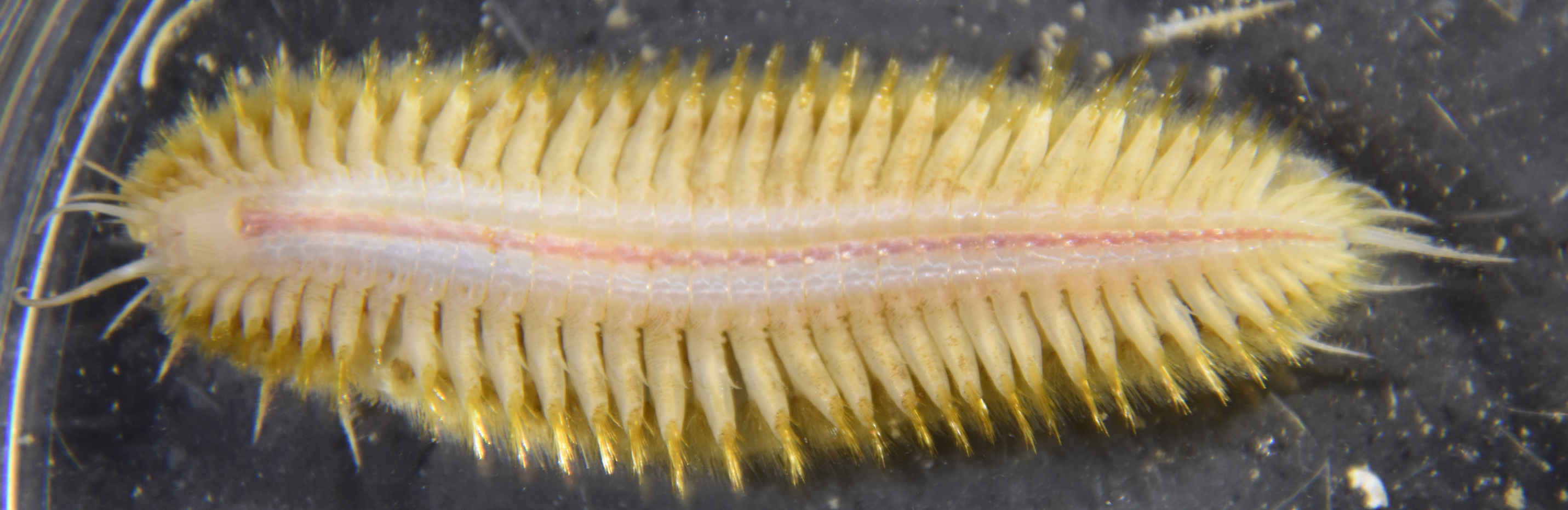
This view of the ventral
side (head to left) shows the individual segments under the elytra.
The amber-colored neurosetae
are most evident on each parapodial
segment, while the thinner capillary
notosetae
are visible
behind (dorsal
to) them.
Photo by Dave Cowles, July 2023
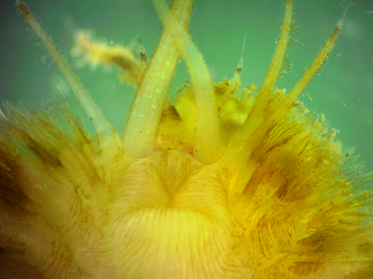
This ventral
view
of the anterior
end
shows the medial antennae
projecting forward (upward in the photo) from the prostomium,
with the lateral
antennae
based just
ventral
to
them. The visible mouth is on the peristomium,
which is the segment immediately behind the prostomium.
To the right, the cluster of thick, amber, bristle-like neurosetae
are seen, while dorsal
to them (above and behind the neurosetae
in the photo) are the thinner, capillary
notosetae.
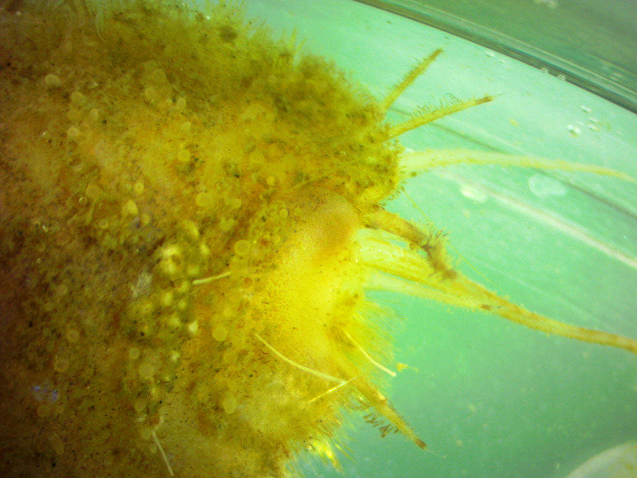
This dorsal
view
of the anterior
end
(facing right) shows the several sets of prostomial
antennae.
The right
front elytra
has dropped
off but the bumps and tubercles
along the other elytra,
especially along their posterior
margins, can be seen. Photo by Dave Cowles, July 2023.
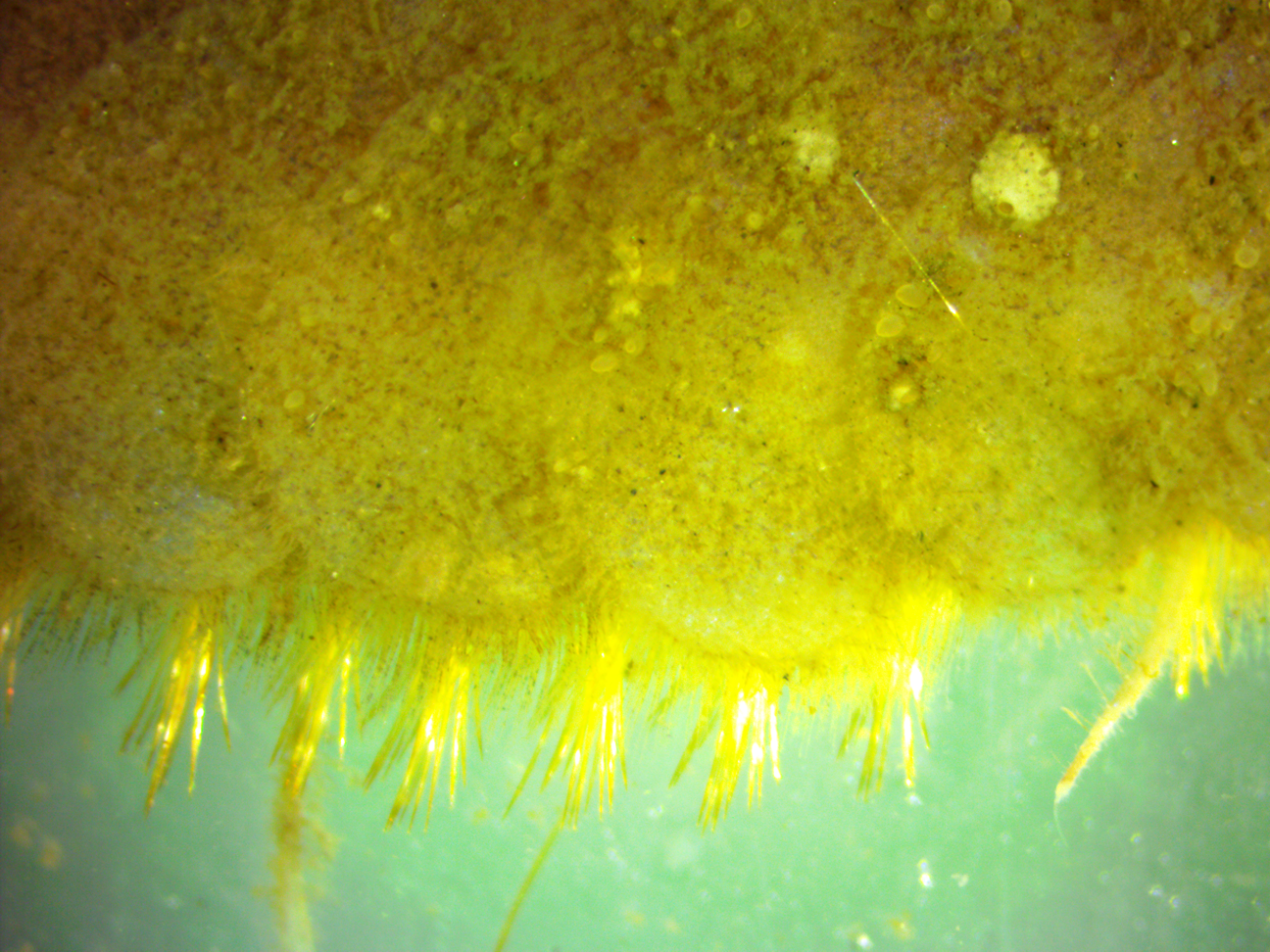
This dorsal
closeup
view of the animal's side shows the fine notosetae
and coarser amber neurosetae
behind (ventral
to)
them projecting from each parapodium.
The textured surface of the elytra
is also visible. Anterior
is to the right. Photo by Dave Cowles, July 2023.
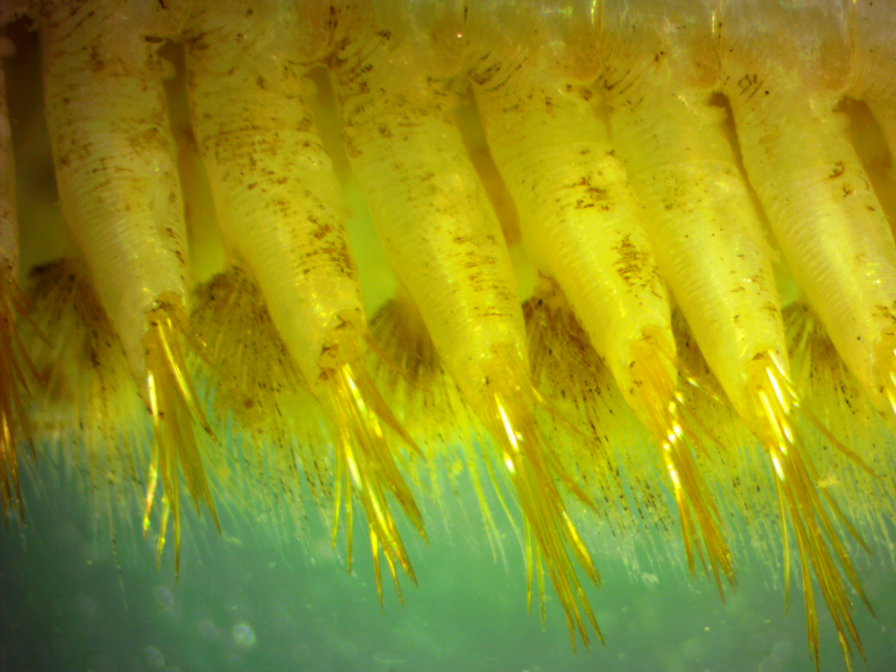
This ventral
view
shows the individual parapodia
and a closer view of the neurosetae,
with the smaller capillary
notosetae
behind
them. Photo by Dave Cowles, July 2023.
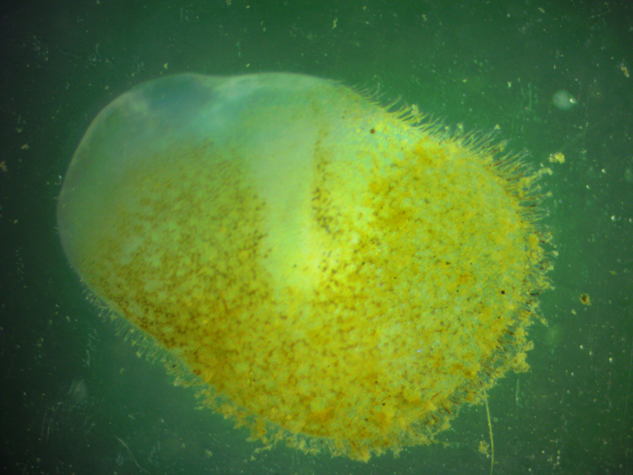
This dorsal
view
of a detached elytra
shows the smooth anterior
surface (upper in this photo) where it was fitted under the elytra
anterior
to it, along
with the bumpy texture and fringing papillae
along the exposed surface. Photo by Dave Cowles, July 2023.
Authors and Editors
of Page:
Dave Cowles (2023): Created original page
CSS coding for page developed by Jonathan Cowles
Salish Sea Invertebrates web site provided courtesy of Walla
Walla University