Paralvinella (Paralvinella) palmiformis Desbruyeres and Laubier, 1986Common name(s): |
|
| Synonyms: | 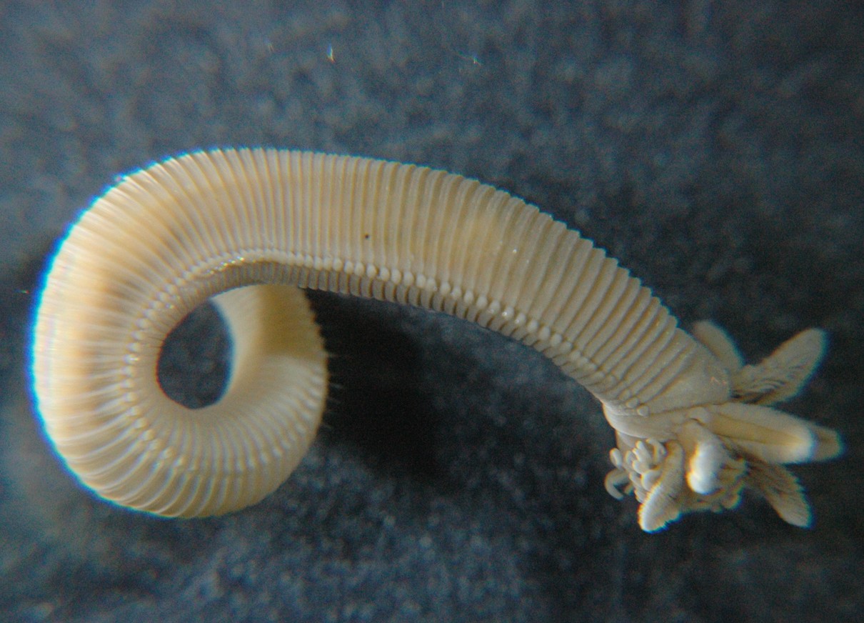 |
|
Phylum Annelida
Subclass Palpata
Order Canalipalpata
Suborder Terebellida
Family Alvinellidae
|
|
| Paralvinella palmiformis, preserved specimen, Collected by Kirt Onthank from hydrothermal vent on Endeavor Ridge. About 3.5 cm long. | |
| (Photo by: Dave Cowles) | |
Description: Order Canalipalpata are the bristle-footed Annelids or fan-head worms. They have no jaws or teeth. Most have grooved, ciliated tentacles with which they feed. Terebellids are mostly sessile tubeworms. Family Alvinellidae is found in the deep sea at hydrothermal vents. They usually build mucus tubes and feed with flattened, ciliated tentacles, plus obtain nutrition from episymbiotic bacteria living on their surface. Paralvinella palmiformis has a body with 100-118 segments, which gradually taper posteriorly (see photo above). The prostomium is reduced in its middle portion. A set of many smooth, grooved tentacles forms the buccal apparatus. Males also have two robust peribuccal tentacles that end in three rounded lobes bordered with papillae, and two blind cavities on the ventral side of the peristomium. The featherlike branchiae (gills) fan out anteriorly, dorsal to the feeding tentacles. The first 20-31 chaetigerous segments have only notopodia (no neuropodia), and segment 7 is highly modified. Color pinkish when preserved in alcohol, brownish red in life. Length up to 8 cm.
How to Distinguish from Similar Species: Of other species that may be found at Pacific Northwest hydrothermal vents, P. pandorae has only about 60 chaetigerous segments and only the first 3 chaetigerous segments have only notopodia. It is light brown to pink in alcohol. P. sulfincola has only about 54-68 chaetigerous segments, and the first 24-30 have only notopodia. Its body doesn't taper much until the last 10 segments. It may turn chocolate brown when preserved in formalin.
Geographical Range: Deep-sea hydrothermal vents in the NE Pacific: Gorda Ridge, Explorer Ridge, Juan de Fuca Ridge
Depth Range: Deep sea, about 2000 m
Habitat: The sulfide chimneys of hydrothermal vents.
Biology/Natural History: Typically live with caudal end coiled around other worm tubes or attached to the surface of sulfide chimneys. They are a deposit feeder and cover themselves with mucus. Paralvinellids also are symbiotic with ectosymbiotic bacteria which live on the mucus. Can withstand rather high temperatures (up to 45 C, Rinke and Lee 2009), but not as high as its congener P. sulfincola can. At its highest temperatures it increases heat-shock protein levels (Dilly et al., 2012). Glycine is the major osmolyte, and it contains less thiotaurine, which may be used in sulphide detoxification, than does P. sulfincola. The species contained no sarcosine (Yancy et al., 2009). Aerobic metabolism appears to dominate in the gills, while anaerobic metabolism (glycolosys) appears to be more prominent in the body wall (Rinke and Lee, 2009).
Wang et al. (2025) discovered in the closely-related species Paralvinella hessleri,
which occurs on hydrothermal vents in the western Pacific, that the
animal has a unique method for detoxifying poisons in its extreme vent
environment. They discovered that the animals sequestered large amounts
of toxic arsenic within their epithelial tissues, comprising up to 1%
of their total wet weight. Further analysis showed that this
arsenic was deposited intracellularly in the form of yellow granules of
an arsenic sulfide called orpiment(As 2S3), thus
"fighting poison with poison" and converting both the toxic arsenic and
toxic sulfide in the vent water into a non-toxic compound. Given the
fact that P. palmaeformis
also generally has a yellow hue, it is very likely that this species
also uses this detoxification method. The red hemoglobin from the blood
along with the yellow orpiment in their epithelium may be responsible
for the orange coloration seen as well. See also Jiang,
Science, October 16, 2025 p. 253 for a summary and photo.
| Return to: | |||
| Main Page | Alphabetic Index | Systematic Index | Glossary |
References:
Dichotomous Keys:General References:
Scientific Articles:
Dilly, Geoffrye F., C. Robert Young, William S. Lane, Jasmyn Pangilinan, and Peter R. Girguis, 2012. Exploring the limit of metazoan thermal tolerance via comparative proteomics: thermally induced changes in protein abundance by two hydrothermal vent polychaetes. Proceedings of the Royal Society B-Biological Sciences 279:1741 pp 3347-3356
Juniper, S. Kim, and Pascale Martineu, 1995. Alvinellids and sulfides at hydrothermal vents of the eastern Pacific: A review. American Zoologist 35:2 pp 174-185
Rinke, C. and R.W. Lee, 2009. Pathways, activities, and thermal stability of anaerobic and aerobic enzymes in thermophilic vent paralvinellid worms. Marine Ecology Progress Series 382: pp 99-112
Wang H, Cao L, Zhang H, Zhong Z, Zhou L, Lian C, et al., 2025. A deep-sea hydrothermal vent worm detoxifies arsenic and sulfur by intracellular biomineralization of orpiment (As2S3). PLoS Biol 23(8): e3003291. https://doi.org/10.1371/journal.pbio.3003291
Yancey, Paul H., Joanna Ishikawa, Brigitte Meyer, Peter Girguis, and Raymond W. Lee, 2009. Thiotaurine and hypotaurine contents in hydrothermal-vent polychaetes without thiotrophic endosymbionts: correlation with sulfide exposure. Journal of Experimental Zoology Part A-Ecological Genetics and Physiology 311A:6 pp 439-447
Web sites:
General Notes and
Observations: Locations,
abundances, unusual behaviors:
My thanks to Kirt Onthank for providing me the preserved specimens photographed on this page.
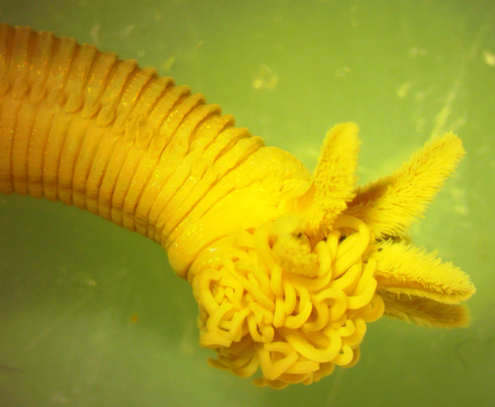
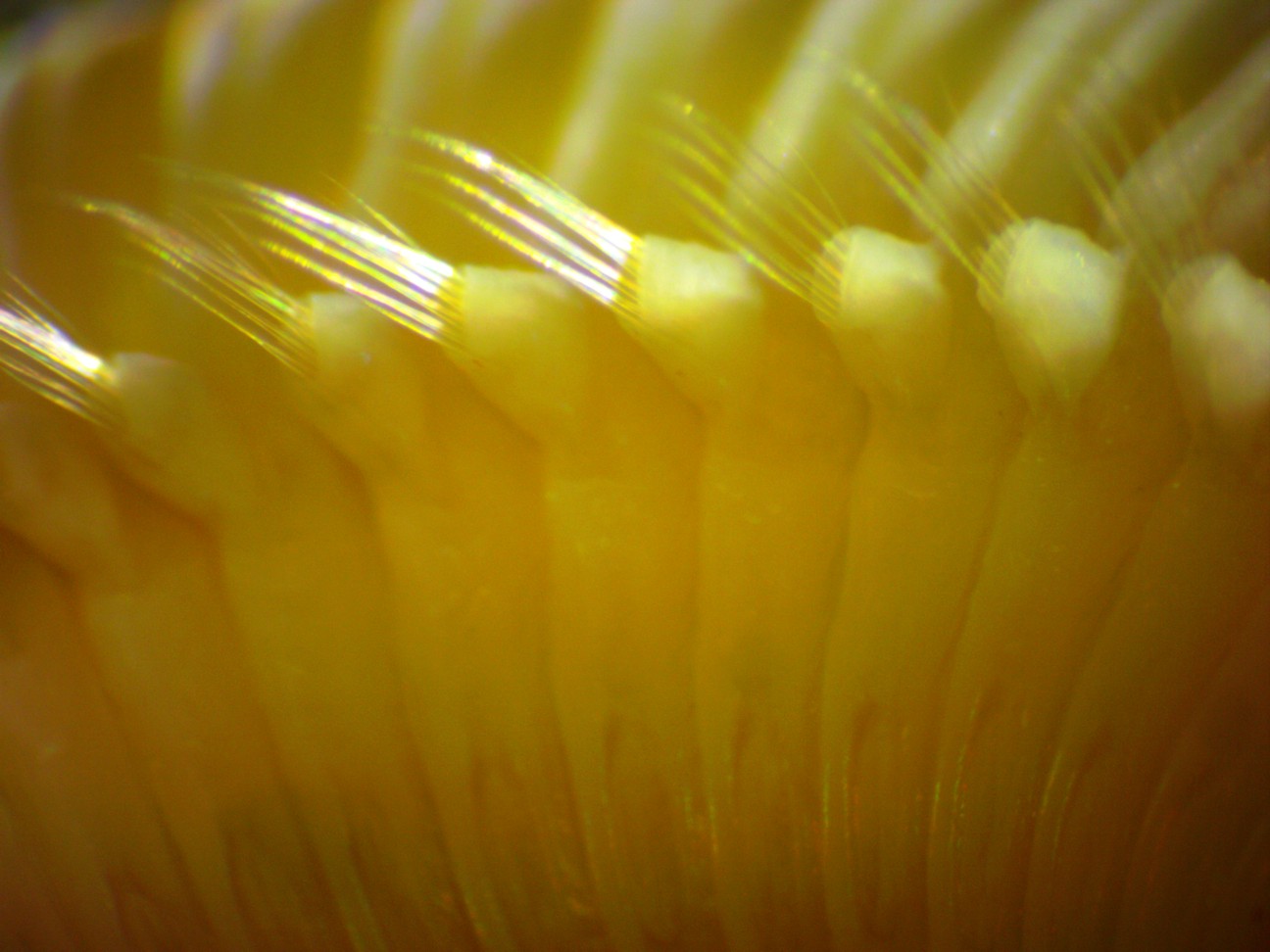
This view of the head of a preserved specimen shows the
spaghetti-like
ciliated tentacles
used for feeding, plus the bushy gills dorsal to them. In
life the
animal would be
bright pink or brownish-red.. It can be difficult for the
non-expert
to determine which side is dorsal and which side is ventral in
polychaetes.
In this case,
the spaghetti-like feeding tentacles are on the ventral side and the
feather-like gills are dorsal.
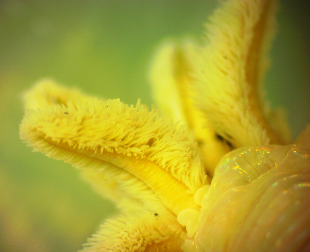
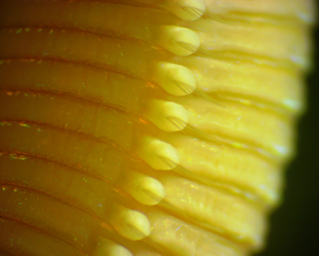
A view of the dorsal (gill) side of the head. The
first chaetigerous
segment (setiger), with a parapodium
and notosetae
projecting,
can be seen near the bottom center. A small amount of debris
has
become attached to the tops of the
notosetae.
Note that the tuft of setae
are on a well-defined (dorsal, notopodial)
lobe of the parapodium
and there is no corresponding ventral lobe (neuropodium)
with neurosetae--this
is one of
the identifying characteristics on the first 20-31 setigers
for this species.
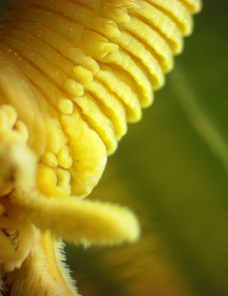
In this view of the first several segments, dorsal is right,
ventral
is left, and the head is downward. The bumps along the sides
of the segments are the parapodia.
Setae
are projecting from the parapodia
although they cannot be seen clearly on all the segments.
The first chaetigerous
segment (setiger)
is
partly hidden behind the head and gill structures. The parapodia
and setae
on setigers
2 and 3 are clearly visible. Chaetigerous
segment 4 is swollen on thedorsal side and at that point the location
of
the parapodia
shifts in the ventral direction. Setae
can be seen projecting from several segments. Note that the parapodium
and setae on setifer 7
are not evident. The species description says that the setae
are much modified on setiger
7. If this modification is like that seen
in other paralvinellids such as P.
sulfincola, the setae are much larger, thicker,
darker, and curved
backward (photo).
Also note the complete lack of a ventral (neuropodium)
lobe on the parapodia
and the lack of neurosetae.
This view shows the parapodia
on the anterior portion of the body. Ventral is to the
left.
The well-
developed dorsal parapodial
lobes (notopodia) with long notosetae
are clearly visible. No ventral
lobes (neuropodia)
nor neurosetae
are
visible.
This ventral
This closeup of the parapodia
on a posterior section of the body shows the well-developed notopodium
(dorsal portion)
with well-developed notosetae.
The ventral portion of the parapodium
(neuropodium,
top
of photo) appears to
be simply a flattened ridge with no neurosetae
visible such as was seen on the anterior portion, although the
neuropodial
ridge is better developed. The species description states
that neurosetae
are present
on the posterior portion of the body so they should be visible
here.
However, in Terebellids, which are other members
of this Order, the neurosetae
are actually very short uncini
which are hard to see. That may be the case here as well.
Authors and Editors of Page:
Dave Cowles (2014): Created original page
CSS coding for page developed by Jonathan Cowles (2007)
Salish Sea Invertebrates web site provided courtesy of Walla
Walla University